

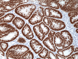
Immunohistochemical staining of formalin fixed and paraffin embedded human colon cancer tissue section using anti-SATB2 rabbit monoclonal antibody (Clone RM365) at a 1:1000 dilution.
Immunohistochemical staining of formalin fixed and paraffin embedded human colon cancer tissue section using anti-SATB2 rabbit monoclonal antibody (Clone RM365) at a 1:1000 dilution.
anti-SATB2 (human), Rabbit Monoclonal (RM365)
REV-31-1251-00
ApplicationsWestern Blot, ImmunoHistoChemistry
Product group Antibodies
ReactivityHuman
TargetSATB2
Overview
- SupplierRevMAb Biosciences
- Product Nameanti-SATB2 (human), Rabbit Monoclonal (RM365)
- Delivery Days Customer2
- ApplicationsWestern Blot, ImmunoHistoChemistry
- CertificationResearch Use Only
- ClonalityMonoclonal
- Clone IDRM365
- Gene ID23314
- Target nameSATB2
- Target descriptionSATB homeobox 2
- Target synonymsC2DELq32q33, DEL2Q32Q33, GLSS, DNA-binding protein SATB2, SATB family member 2, SATB2 fusion, special AT-rich sequence-binding protein 2
- HostRabbit
- IsotypeIgG
- Protein IDQ9UPW6
- Protein NameDNA-binding protein SATB2
- Scientific DescriptionDNA-binding protein SATB2 binds to DNA at the nuclear matrix- or scaffold associated regions. It is thought to recognize the sugar-phosphate structure of double-stranded DNA. SATB2 is a transcription factor controlling nuclear gene expression by binding to matrix attachment regions (MARs) of DNA and inducing a local chromatin-loop remodeling. It also acts as a docking site for several chromatin remodeling enzymes by recruiting corepressors (HDACs) or coactivators (HATs) directly to promoters and enhancers. SATB2 is required for the initiation of the upper-layer neurons (UL1) specific genetic program and for the inactivation of deep-layer neurons (DL) and UL2 specific genes, probably by modulating BCL11B expression. It is a repressor of Ctip2 and a regulatory determinant of corticocortical connections in the developing cerebral cortex. Mutations in the gene are associate with cleft palate isolated (CPI). SATB2 expression is highly specific for cancer in the lower GI-tract and has been implicated as a cancer biomarker for colorectal cancer. - Recombinant Antibody. This antibody reacts human DNA-binding protein SATB2. Applications: WB, IHC. Source: Rabbit. Liquid. 50% Glycerol/PBS with 1% BSA and 0.09% sodium azide. DNA-binding protein SATB2 binds to DNA at the nuclear matrix- or scaffold associated regions. It is thought to recognize the sugar-phosphate structure of double-stranded DNA. SATB2 is a transcription factor controlling nuclear gene expression by binding to matrix attachment regions (MARs) of DNA and inducing a local chromatin-loop remodeling. It also acts as a docking site for several chromatin remodeling enzymes by recruiting corepressors (HDACs) or coactivators (HATs) directly to promoters and enhancers. SATB2 is required for the initiation of the upper-layer neurons (UL1) specific genetic program and for the inactivation of deep-layer neurons (DL) and UL2 specific genes, probably by modulating BCL11B expression. It is a repressor of Ctip2 and a regulatory determinant of corticocortical connections in the developing cerebral cortex. Mutations in the gene are associate with cleft palate isolated (CPI). SATB2 expression is highly specific for cancer in the lower GI-tract and has been implicated as a cancer biomarker for colorectal cancer.
- ReactivityHuman
- Storage Instruction-20°C,2°C to 8°C
- UNSPSC41116161
Datasheet
Related products
Product group Antibodies
Anti-SATB2 AntibodyA91312
ApplicationsWestern Blot, ImmunoHistoChemistry
ReactivityHuman, Rat
- SizePrice

Product group Antibodies
Anti-SATB2 Antibody Picoband(r)A02588-2-CARRIER-FREE
ApplicationsWestern Blot
ReactivityHuman
TargetSATB2
- SizePrice
Product group Antibodies
Anti-SATB2 Antibody144-64612
ApplicationsWestern Blot
ReactivityHuman
TargetSATB2
- SizePrice

Product group Antibodies
Anti-SATB2 AntibodyAMAB90678
ApplicationsWestern Blot, ImmunoHistoChemistry
ReactivityHuman, Rat
TargetSATB2
- SizePrice
Product group Antibodies
References
SATB2 Polyclonal AntibodyBS-11949R
ApplicationsFlow Cytometry, ImmunoFluorescence, Western Blot, ELISA, ImmunoCytoChemistry, ImmunoHistoChemistry, ImmunoHistoChemistry Frozen, ImmunoHistoChemistry Paraffin
ReactivityBovine, Equine, Human, Mouse, Rat, Sheep
TargetSATB2
- SizePrice
Product group Antibodies
SATB2 AntibodyCSB-PA892170LA01HU
ApplicationsELISA, ImmunoHistoChemistry
ReactivityHuman
TargetSATB2
- SizePrice
Product group Antibodies
ApplicationsImmunoPrecipitation, Western Blot, ImmunoCytoChemistry, ImmunoHistoChemistry
TargetSATB2
- SizePrice

Product group Antibodies
SATB2 antibodyGTX132972
ApplicationsImmunoFluorescence, Western Blot, ImmunoCytoChemistry
ReactivityHuman, Rat
TargetSATB2
- SizePrice
Product group Antibodies

SATB2 Antibody (aa250-300)LS-C288640
ApplicationsImmunoPrecipitation
ReactivityHuman
TargetSATB2
- SizePrice