

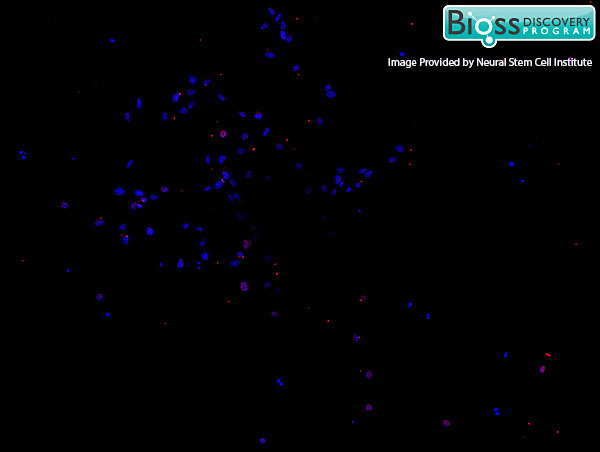
Mouse embryonic cortex cells labeled with RABBIT ANTI-SATB2 POLYCLONAL ANTIBODY Polyclonal Antibody, conjugated (bs-11949R-Cy3) followed by DAPI staining.
 at 1:200, followed by conjugation to the secondary antibody and DAB staining\n")
Mouse embryonic cortex cells labeled with RABBIT ANTI-SATB2 POLYCLONAL ANTIBODY Polyclonal Antibody, conjugated (bs-11949R-Cy3) followed by DAPI staining.
SATB2 Polyclonal Antibody
BS-11949R
ApplicationsFlow Cytometry, ImmunoFluorescence, Western Blot, ELISA, ImmunoCytoChemistry, ImmunoHistoChemistry, ImmunoHistoChemistry Frozen, ImmunoHistoChemistry Paraffin
Product group Antibodies
ReactivityBovine, Equine, Human, Mouse, Rat, Sheep
TargetSATB2
Overview
- SupplierBioss
- Product NameSATB2 Polyclonal Antibody
- Delivery Days Customer16
- ApplicationsFlow Cytometry, ImmunoFluorescence, Western Blot, ELISA, ImmunoCytoChemistry, ImmunoHistoChemistry, ImmunoHistoChemistry Frozen, ImmunoHistoChemistry Paraffin
- Applications SupplierWB(1:300-5000), ELISA(1:500-1000), FCM(1:20-100), IHC-P(1:200-400), IHC-F(1:100-500), IF(IHC-P)(1:50-200), IF(IHC-F)(1:50-200), IF(ICC)(1:50-200)
- CertificationResearch Use Only
- ClonalityPolyclonal
- Concentration1 ug/ul
- ConjugateUnconjugated
- Gene ID23314
- Target nameSATB2
- Target descriptionSATB homeobox 2
- Target synonymsC2DELq32q33, DEL2Q32Q33, GLSS, DNA-binding protein SATB2, SATB family member 2, SATB2 fusion, special AT-rich sequence-binding protein 2
- HostRabbit
- IsotypeIgG
- Protein IDQ9UPW6
- Protein NameDNA-binding protein SATB2
- ReactivityBovine, Equine, Human, Mouse, Rat, Sheep
- Storage Instruction-20°C
- UNSPSC41116161
References
- Skeletal impact of 17beta-estradiol in T cell-deficient mice: age-dependent bone effects and osteosarcoma formation. Cheng JN et al., 2020 Apr, Clin Exp MetastasisRead this paper
Datasheet
Related products
Product group Antibodies
Anti-SATB2 AntibodyA91312
ApplicationsWestern Blot, ImmunoHistoChemistry
ReactivityHuman, Rat
- SizePrice

Product group Antibodies
Anti-SATB2 Antibody Picoband(r)A02588-2-CARRIER-FREE
ApplicationsWestern Blot
ReactivityHuman
TargetSATB2
- SizePrice
Product group Antibodies
Anti-SATB2 Antibody144-64612
ApplicationsWestern Blot
ReactivityHuman
TargetSATB2
- SizePrice

Product group Antibodies
Anti-SATB2 AntibodyAMAB90678
ApplicationsWestern Blot, ImmunoHistoChemistry
ReactivityHuman, Rat
TargetSATB2
- SizePrice
Product group Antibodies
SATB2 AntibodyCSB-PA892170LA01HU
ApplicationsELISA, ImmunoHistoChemistry
ReactivityHuman
TargetSATB2
- SizePrice
Product group Antibodies
ApplicationsImmunoPrecipitation, Western Blot, ImmunoCytoChemistry, ImmunoHistoChemistry
TargetSATB2
- SizePrice

Product group Antibodies
SATB2 antibodyGTX132972
ApplicationsImmunoFluorescence, Western Blot, ImmunoCytoChemistry
ReactivityHuman, Rat
TargetSATB2
- SizePrice
Product group Antibodies

SATB2 Antibody (aa250-300)LS-C288640
ApplicationsImmunoPrecipitation
ReactivityHuman
TargetSATB2
- SizePrice