


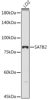
Various whole cell extracts (30 μg) were separated by 7.5% SDS-PAGE, and the membrane was blotted with SATB2 antibody (GTX132972) diluted at 1:1000. The HRP-conjugated anti-rabbit IgG antibody (GTX213110-01) was used to detect the primary antibody. Corresponding RNA expression data for the same cell lines are based on Human Protein Atlas program.
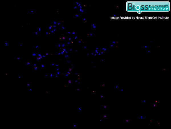
![SATB2 antibody detects SATB2 protein by immunofluorescent analysis. Sample: DIV10 rat E18 primary cortical neuron cells were fixed in 4% paraformaldehyde at RT for 15 min. Green: SATB2 stained by SATB2 antibody (GTX132972) diluted at 1:500. Red: Tau, stained by Tau antibody [GT287] (GTX634809) diluted at 1:500. Blue: Fluoroshield with DAPI (GTX30920).](https://www.genetex.com/upload/website/prouct_img/normal/GTX132972/GTX132972_43551_20191125_ICC_IF_R_w_23060523_728.webp "SATB2 antibody detects SATB2 protein by immunofluorescent analysis. Sample: DIV10 rat E18 primary cortical neuron cells were fixed in 4% paraformaldehyde at RT for 15 min. Green: SATB2 stained by SATB2 antibody (GTX132972) diluted at 1:500. Red: Tau, stained by Tau antibody [GT287] (GTX634809) diluted at 1:500. Blue: Fluoroshield with DAPI (GTX30920).")
Various whole cell extracts (30 μg) were separated by 7.5% SDS-PAGE, and the membrane was blotted with SATB2 antibody (GTX132972) diluted at 1:1000. The HRP-conjugated anti-rabbit IgG antibody (GTX213110-01) was used to detect the primary antibody. Corresponding RNA expression data for the same cell lines are based on Human Protein Atlas program.
SATB2 antibody
GTX132972
ApplicationsImmunoFluorescence, Western Blot, ImmunoCytoChemistry
Product group Antibodies
ReactivityHuman, Rat
TargetSATB2
Overview
- SupplierGeneTex
- Product NameSATB2 antibody
- Delivery Days Customer9
- Application Supplier NoteWB: 1:500-1:3000. *Optimal dilutions/concentrations should be determined by the researcher.Not tested in other applications.
- ApplicationsImmunoFluorescence, Western Blot, ImmunoCytoChemistry
- CertificationResearch Use Only
- ClonalityPolyclonal
- Concentration1.34 mg/ml
- ConjugateUnconjugated
- Gene ID23314
- Target nameSATB2
- Target descriptionSATB homeobox 2
- Target synonymsC2DELq32q33, DEL2Q32Q33, GLSS, DNA-binding protein SATB2, SATB family member 2, SATB2 fusion, special AT-rich sequence-binding protein 2
- HostRabbit
- IsotypeIgG
- Protein IDQ9UPW6
- Protein NameDNA-binding protein SATB2
- Scientific DescriptionThis gene encodes a DNA binding protein that specifically binds nuclear matrix attachment regions. The encoded protein is involved in transcription regulation and chromatin remodeling. Defects in this gene are associated with isolated cleft palate and cognitive disability. Alternate splicing results in multiple transcript variants that encode the same protein. [provided by RefSeq, Feb 2010]
- ReactivityHuman, Rat
- Storage Instruction-20°C or -80°C,2°C to 8°C
- UNSPSC41116161
Datasheet
Related products
Product group Antibodies
Anti-SATB2 AntibodyA91312
ApplicationsWestern Blot, ImmunoHistoChemistry
ReactivityHuman, Rat
- SizePrice

Product group Antibodies
Anti-SATB2 Antibody Picoband(r)A02588-2-CARRIER-FREE
ApplicationsWestern Blot
ReactivityHuman
TargetSATB2
- SizePrice
Product group Antibodies
Anti-SATB2 Antibody144-64612
ApplicationsWestern Blot
ReactivityHuman
TargetSATB2
- SizePrice

Product group Antibodies
Anti-SATB2 AntibodyAMAB90678
ApplicationsWestern Blot, ImmunoHistoChemistry
ReactivityHuman, Rat
TargetSATB2
- SizePrice
Product group Antibodies
References
SATB2 Polyclonal AntibodyBS-11949R
ApplicationsFlow Cytometry, ImmunoFluorescence, Western Blot, ELISA, ImmunoCytoChemistry, ImmunoHistoChemistry, ImmunoHistoChemistry Frozen, ImmunoHistoChemistry Paraffin
ReactivityBovine, Equine, Human, Mouse, Rat, Sheep
TargetSATB2
- SizePrice
Product group Antibodies
SATB2 AntibodyCSB-PA892170LA01HU
ApplicationsELISA, ImmunoHistoChemistry
ReactivityHuman
TargetSATB2
- SizePrice
Product group Antibodies
ApplicationsImmunoPrecipitation, Western Blot, ImmunoCytoChemistry, ImmunoHistoChemistry
TargetSATB2
- SizePrice
Product group Antibodies
SATB2 antibodyGTX30894
ApplicationsWestern Blot, ELISA, ImmunoHistoChemistry, ImmunoHistoChemistry Paraffin
ReactivityHuman, Mouse, Rat
TargetSATB2
- SizePrice
Product group Antibodies
SATB2 antibodyGTX31773
ApplicationsWestern Blot, ELISA, ImmunoHistoChemistry, ImmunoHistoChemistry Paraffin
ReactivityHuman, Mouse, Rat
TargetSATB2
- SizePrice