


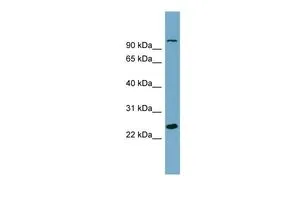
Figure 1. Western blot analysis of SEMA3D using anti-SEMA3D antibody (A09239-1). Electrophoresis was performed on a 5-20% SDS-PAGE gel at 70V (Stacking gel) / 90V (Resolving gel) for 2-3 hours. The sample well of each lane was loaded with 30 ug of sample under reducing conditions. Lane 1: human Hacat whole cell lysates, Lane 2: human PC-3 whole cell lysates, Lane 3: human 293T whole cell lysates, Lane 4: human U20S whole cell lysates. After electrophoresis, proteins were transferred to a nitrocellulose membrane at 150 mA for 50-90 minutes. Blocked the membrane with 5% non-fat milk/TBS for 1.5 hour at RT. The membrane was incubated with rabbit anti-SEMA3D antigen affinity purified polyclonal antibody (Catalog # A09239-1) at 0.5 microg/mL overnight at 4°C, then washed with TBS-0.1%Tween 3 times with 5 minutes each and probed with a goat anti-rabbit IgG-HRP secondary antibody at a dilution of 1:5000 for 1.5 hour at RT. The signal is developed using an Enhanced Chemiluminescent detection (ECL) kit (Catalog # EK1002) with Tanon 5200 system. A specific band was detected for SEMA3D at approximately 90 kDa. The expected band size for SEMA3D is at 88,90 kDa.
Figure 1. Western blot analysis of SEMA3D using anti-SEMA3D antibody (A09239-1). Electrophoresis was performed on a 5-20% SDS-PAGE gel at 70V (Stacking gel) / 90V (Resolving gel) for 2-3 hours. The sample well of each lane was loaded with 30 ug of sample under reducing conditions. Lane 1: human Hacat whole cell lysates, Lane 2: human PC-3 whole cell lysates, Lane 3: human 293T whole cell lysates, Lane 4: human U20S whole cell lysates. After electrophoresis, proteins were transferred to a nitrocellulose membrane at 150 mA for 50-90 minutes. Blocked the membrane with 5% non-fat milk/TBS for 1.5 hour at RT. The membrane was incubated with rabbit anti-SEMA3D antigen affinity purified polyclonal antibody (Catalog # A09239-1) at 0.5 microg/mL overnight at 4°C, then washed with TBS-0.1%Tween 3 times with 5 minutes each and probed with a goat anti-rabbit IgG-HRP secondary antibody at a dilution of 1:5000 for 1.5 hour at RT. The signal is developed using an Enhanced Chemiluminescent detection (ECL) kit (Catalog # EK1002) with Tanon 5200 system. A specific band was detected for SEMA3D at approximately 90 kDa. The expected band size for SEMA3D is at 88,90 kDa.
Anti-SEMA3D Antibody Picoband(r)
A09239-1-CARRIER-FREE
ApplicationsWestern Blot, ELISA
Product group Antibodies
ReactivityHuman
TargetSEMA3D
Overview
- SupplierBoster Bio
- Product NameAnti-SEMA3D Antibody Picoband(r)
- Delivery Days Customer9
- ApplicationsWestern Blot, ELISA
- CertificationResearch Use Only
- ClonalityPolyclonal
- Concentration500 ug/ml
- Gene ID223117
- Target nameSEMA3D
- Target descriptionsemaphorin 3D
- Target synonymsSema-Z2, coll-2, semaphorin-3D, collapsin 2, sema domain, immunoglobulin domain (Ig), short basic domain, secreted, (semaphorin) 3D
- HostRabbit
- IsotypeIgG
- Protein IDO95025
- Protein NameSemaphorin-3D
- Scientific DescriptionBoster Bio Anti-SEMA3D Antibody Picoband® catalog # A09239-1. Tested in ELISA, WB applications. This antibody reacts with Human. The brand Picoband indicates this is a premium antibody that guarantees superior quality, high affinity, and strong signals with minimal background in Western blot applications. Only our best-performing antibodies are designated as Picoband, ensuring unmatched performance.
- ReactivityHuman
- Storage Instruction-20°C,2°C to 8°C
- UNSPSC12352203
Related products
Product group Antibodies
SEMA3D AntibodyCSB-PA065596
ApplicationsELISA, ImmunoHistoChemistry
ReactivityHuman, Mouse
TargetSEMA3D
- SizePrice
Product group Antibodies
Anti-SEMA3D AntibodyA45529
ApplicationsImmunoHistoChemistry
ReactivityHuman, Mouse
- SizePrice

Product group Antibodies
Anti-SEMA3D AntibodyHPA037522
ApplicationsImmunoHistoChemistry
ReactivityHuman
TargetSEMA3D
- SizePrice
Product group Antibodies

SEMA3D / Semaphorin 3D AntibodyLS-C402780
ApplicationsELISA, ImmunoHistoChemistry
ReactivityHuman, Mouse
TargetSEMA3D
- SizePrice
Product group Antibodies
SEMA3D antibody, InternalGTX45262
ApplicationsWestern Blot
ReactivityHuman
TargetSEMA3D
- SizePrice