

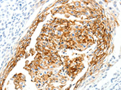
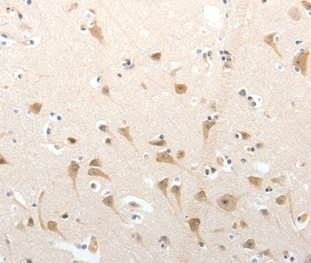
The image on the left is immunohistochemistry of paraffin-embedded Human cervical cancer tissue using CSB-PA065596(SEMA3D Antibody) at dilution 1/40, on the right is treated with synthetic peptide. (Original magnification: x200)
The image on the left is immunohistochemistry of paraffin-embedded Human cervical cancer tissue using CSB-PA065596(SEMA3D Antibody) at dilution 1/40, on the right is treated with synthetic peptide. (Original magnification: x200)
SEMA3D Antibody
CSB-PA065596
ApplicationsELISA, ImmunoHistoChemistry
Product group Antibodies
ReactivityHuman, Mouse
TargetSEMA3D
Overview
- SupplierCusabio
- Product NameSEMA3D Antibody
- Delivery Days Customer20
- ApplicationsELISA, ImmunoHistoChemistry
- CertificationResearch Use Only
- ClonalityPolyclonal
- ConjugateUnconjugated
- Gene ID223117
- Target nameSEMA3D
- Target descriptionsemaphorin 3D
- Target synonymsSema-Z2, coll-2, semaphorin-3D, collapsin 2, sema domain, immunoglobulin domain (Ig), short basic domain, secreted, (semaphorin) 3D
- HostRabbit
- IsotypeIgG
- Protein IDO95025
- Protein NameSemaphorin-3D
- Scientific DescriptionThis gene Belongs to the semaphorin family. Induces the collapse and paralysis of neuronal growth cones. Could potentially act as repulsive cues toward specific neuronal populations. Strong binding to neuropilin is mediated by the carboxy third of the protein.
- ReactivityHuman, Mouse
- Storage Instruction-20°C or -80°C
- UNSPSC41116161
Related products

Product group Antibodies
Anti-SEMA3D Antibody Picoband(r)A09239-1-CARRIER-FREE
ApplicationsWestern Blot, ELISA
ReactivityHuman
TargetSEMA3D
- SizePrice
Product group Antibodies
Anti-SEMA3D AntibodyA45529
ApplicationsImmunoHistoChemistry
ReactivityHuman, Mouse
- SizePrice

Product group Antibodies
Anti-SEMA3D AntibodyHPA037522
ApplicationsImmunoHistoChemistry
ReactivityHuman
TargetSEMA3D
- SizePrice
Product group Antibodies

SEMA3D / Semaphorin 3D AntibodyLS-C402780
ApplicationsELISA, ImmunoHistoChemistry
ReactivityHuman, Mouse
TargetSEMA3D
- SizePrice
Product group Antibodies
SEMA3D antibody, InternalGTX45262
ApplicationsWestern Blot
ReactivityHuman
TargetSEMA3D
- SizePrice