

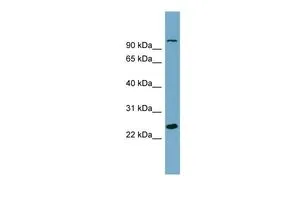
WB analysis of THP-1 cells using GTX45262 SEMA3D antibody at 0.2-1μg/ml.
WB analysis of THP-1 cells using GTX45262 SEMA3D antibody at 0.2-1μg/ml.
SEMA3D antibody, Internal
GTX45262
ApplicationsWestern Blot
Product group Antibodies
ReactivityHuman
TargetSEMA3D
Overview
- SupplierGeneTex
- Product NameSEMA3D antibody, Internal
- Delivery Days Customer9
- Application Supplier NoteWB: 0.2-2.5 ug/ml. *Optimal dilutions/concentrations should be determined by the researcher.Not tested in other applications.
- ApplicationsWestern Blot
- CertificationResearch Use Only
- ClonalityPolyclonal
- Concentration0.5-1 mg/ml
- ConjugateUnconjugated
- Gene ID223117
- Target nameSEMA3D
- Target descriptionsemaphorin 3D
- Target synonymsSema-Z2, coll-2, semaphorin-3D, collapsin 2, sema domain, immunoglobulin domain (Ig), short basic domain, secreted, (semaphorin) 3D
- HostRabbit
- IsotypeIgG
- Protein IDO95025
- Protein NameSemaphorin-3D
- Scientific DescriptionThis gene encodes a member of the semaphorin III family of secreted signaling proteins that are involved in axon guidance during neuronal development. The encoded protein contains an N-terminal Sema domain, an immunoglobulin like domain and a C-terminal basic domain. The protein encoded by this gene binds neuropilin and plays an important role in cardiovascular development. [provided by RefSeq, Aug 2016]
- ReactivityHuman
- Storage Instruction-20°C or -80°C,2°C to 8°C
- UNSPSC41116161
Datasheet
Related products
Product group Antibodies
SEMA3D AntibodyCSB-PA065596
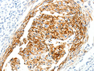
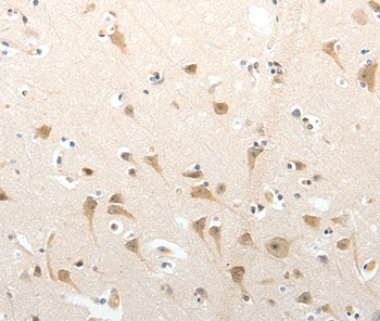
ApplicationsELISA, ImmunoHistoChemistry
ReactivityHuman, Mouse
TargetSEMA3D
- SizePrice

Product group Antibodies
Anti-SEMA3D Antibody Picoband(r)A09239-1-CARRIER-FREE
ApplicationsWestern Blot, ELISA
ReactivityHuman
TargetSEMA3D
- SizePrice
Product group Antibodies
Anti-SEMA3D AntibodyA45529
ApplicationsImmunoHistoChemistry
ReactivityHuman, Mouse
- SizePrice

Product group Antibodies
Anti-SEMA3D AntibodyHPA037522
ApplicationsImmunoHistoChemistry
ReactivityHuman
TargetSEMA3D
- SizePrice
Product group Antibodies

SEMA3D / Semaphorin 3D AntibodyLS-C402780
ApplicationsELISA, ImmunoHistoChemistry
ReactivityHuman, Mouse
TargetSEMA3D
- SizePrice