


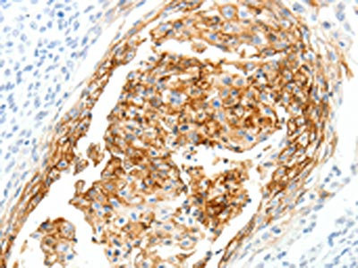
Immunohistochemical staining of human colon shows moderate cytoplasmic positivity in glandular cells.
Immunohistochemical staining of human colon shows moderate cytoplasmic positivity in glandular cells.
Anti-SEMA3D Antibody
HPA037522
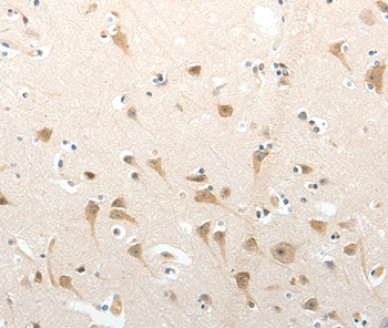
ApplicationsImmunoHistoChemistry
Product group Antibodies
ReactivityHuman
TargetSEMA3D
Overview
- SupplierAtlas Antibodies
- Product NameAnti-SEMA3D Antibody
- Delivery Days Customer4
- ApplicationsImmunoHistoChemistry
- CertificationResearch Use Only
- ClonalityPolyclonal
- ConjugateUnconjugated
- Gene ID223117
- Target nameSEMA3D
- Target descriptionsemaphorin 3D
- Target synonymsSema-Z2, coll-2, semaphorin-3D, collapsin 2, sema domain, immunoglobulin domain (Ig), short basic domain, secreted, (semaphorin) 3D
- HostRabbit
- IsotypeIgG
- Protein IDO95025
- Protein NameSemaphorin-3D
- Scientific DescriptionRecombinant Protein Epitope Signature Tag (PrEST) antigen sequence
- ReactivityHuman
- Storage Instruction-20°C,2°C to 8°C
- UNSPSC41116161
Datasheet
MSDS
Related products
Product group Antibodies
SEMA3D AntibodyCSB-PA065596
ApplicationsELISA, ImmunoHistoChemistry
ReactivityHuman, Mouse
TargetSEMA3D
- SizePrice

Product group Antibodies
Anti-SEMA3D Antibody Picoband(r)A09239-1-CARRIER-FREE
ApplicationsWestern Blot, ELISA
ReactivityHuman
TargetSEMA3D
- SizePrice
Product group Antibodies
Anti-SEMA3D AntibodyA45529
ApplicationsImmunoHistoChemistry
ReactivityHuman, Mouse
- SizePrice
Product group Antibodies

SEMA3D / Semaphorin 3D AntibodyLS-C402780
ApplicationsELISA, ImmunoHistoChemistry
ReactivityHuman, Mouse
TargetSEMA3D
- SizePrice
Product group Antibodies
SEMA3D antibody, InternalGTX45262
ApplicationsWestern Blot
ReactivityHuman
TargetSEMA3D
- SizePrice