


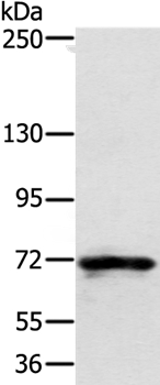
Figure 1. Western blot analysis of Semaphorin 7a/SEMA7A using anti-Semaphorin 7a/SEMA7A antibody (A03832-2). Electrophoresis was performed on a 5-20% SDS-PAGE gel at 70V (Stacking gel) / 90V (Resolving gel) for 2-3 hours. The sample well of each lane was loaded with 30 ug of sample under reducing conditions. Lane 1: human U251 whole cell lysates. After electrophoresis, proteins were transferred to a nitrocellulose membrane at 150 mA for 50-90 minutes. Blocked the membrane with 5% non-fat milk/TBS for 1.5 hour at RT. The membrane was incubated with rabbit anti-Semaphorin 7a/SEMA7A antigen affinity purified polyclonal antibody (Catalog # A03832-2) at 0.5 microg/mL overnight at 4°C, then washed with TBS-0.1%Tween 3 times with 5 minutes each and probed with a goat anti-rabbit IgG-HRP secondary antibody at a dilution of 1:5000 for 1.5 hour at RT. The signal is developed using an Enhanced Chemiluminescent detection (ECL) kit (Catalog # EK1002) with Tanon 5200 system. A specific band was detected for Semaphorin 7a/SEMA7A at approximately 75 kDa. The expected band size for Semaphorin 7a/SEMA7A is at 75 kDa.
. Overlay histogram showing HEL cells stained with A03832-2 (Blue line). To facilitate intracellular staining, cells were fixed with 4% paraformaldehyde and permeabilized with permeabilization buffer. The cells were blocked with 10% normal goat serum. And then incubated with rabbit anti-Semaphorin 7a/SEMA7A Antibody (A03832-2, 1 microg/1x106 cells) for 30 min at 20°C. DyLight®488 conjugated goat anti-rabbit IgG (BA1127, 5-10 microg/1x106 cells) was used as secondary antibody for 30 minutes at 20°C. Isotype control antibody (Green line) was rabbit IgG (1 microg/1x106) used under the same conditions. Unlabelled sample without incubation with primary antibody and secondary antibody (Red line) was used as a blank control.")
Figure 1. Western blot analysis of Semaphorin 7a/SEMA7A using anti-Semaphorin 7a/SEMA7A antibody (A03832-2). Electrophoresis was performed on a 5-20% SDS-PAGE gel at 70V (Stacking gel) / 90V (Resolving gel) for 2-3 hours. The sample well of each lane was loaded with 30 ug of sample under reducing conditions. Lane 1: human U251 whole cell lysates. After electrophoresis, proteins were transferred to a nitrocellulose membrane at 150 mA for 50-90 minutes. Blocked the membrane with 5% non-fat milk/TBS for 1.5 hour at RT. The membrane was incubated with rabbit anti-Semaphorin 7a/SEMA7A antigen affinity purified polyclonal antibody (Catalog # A03832-2) at 0.5 microg/mL overnight at 4°C, then washed with TBS-0.1%Tween 3 times with 5 minutes each and probed with a goat anti-rabbit IgG-HRP secondary antibody at a dilution of 1:5000 for 1.5 hour at RT. The signal is developed using an Enhanced Chemiluminescent detection (ECL) kit (Catalog # EK1002) with Tanon 5200 system. A specific band was detected for Semaphorin 7a/SEMA7A at approximately 75 kDa. The expected band size for Semaphorin 7a/SEMA7A is at 75 kDa.
Anti-Semaphorin 7a/SEMA7A Antibody Picoband(r)
A03832-2-CARRIER-FREE
ApplicationsFlow Cytometry, Western Blot, ELISA
Product group Antibodies
ReactivityHuman
TargetSEMA7A
Overview
- SupplierBoster Bio
- Product NameAnti-Semaphorin 7a/SEMA7A Antibody Picoband(r)
- Delivery Days Customer9
- ApplicationsFlow Cytometry, Western Blot, ELISA
- CertificationResearch Use Only
- ClonalityPolyclonal
- Concentration500 ug/ml
- Gene ID8482
- Target nameSEMA7A
- Target descriptionsemaphorin 7A (JohnMiltonHagen blood group)
- Target synonymsCD108, CDw108, H-SEMA-K1, H-Sema-L, JMH, PFIC11, SEMAK1, SEMAL, semaphorin-7A, JMH blood group antigen, John Milton Hagen blood group H-Sema K1, john-Milton-Hargen human blood group Ag, sema K1, sema L, sema domain, immunoglobulin domain (Ig), and GPI membrane anchor, (semaphorin) 7A (JMH blood group), sema domain, immunoglobulin domain (Ig), and GPI membrane anchor, 7A, semaphorin 7A (John Milton Hagen blood group), semaphorin 7A, GPI membrane anchor (John Milton Hagen blood group), semaphorin-K1, semaphorin-L
- HostRabbit
- IsotypeIgG
- Protein IDO75326
- Protein NameSemaphorin-7A
- Scientific DescriptionBoster Bio Anti-Semaphorin 7a/SEMA7A Antibody Picoband® catalog # A03832-2. Tested in ELISA, Flow Cytometry, WB applications. This antibody reacts with Human. The brand Picoband indicates this is a premium antibody that guarantees superior quality, high affinity, and strong signals with minimal background in Western blot applications. Only our best-performing antibodies are designated as Picoband, ensuring unmatched performance.
- ReactivityHuman
- Storage Instruction-20°C,2°C to 8°C
- UNSPSC12352203
Related products
Product group Antibodies
Anti-SEMA7A AntibodyA37320
ApplicationsWestern Blot, ImmunoHistoChemistry
ReactivityHuman, Mouse
- SizePrice
Product group Antibodies
Anti-SEMA7A Antibody144-62182
ApplicationsWestern Blot
ReactivityHuman
TargetSEMA7A
- SizePrice
Product group Antibodies

SEMA7A / Semaphorin 7A AntibodyLS-C747248
ApplicationsWestern Blot
ReactivityHuman
TargetSEMA7A
- SizePrice
Product group Antibodies
CD108 Polyclonal AntibodyBS-2702R
ApplicationsImmunoFluorescence, Western Blot, ELISA, ImmunoCytoChemistry, ImmunoHistoChemistry, ImmunoHistoChemistry Frozen, ImmunoHistoChemistry Paraffin
ReactivityBovine, Canine, Human, Mouse, Rat
TargetSEMA7A
- SizePrice
Product group Antibodies
SEMA7A AntibodyCSB-PA001418
ApplicationsWestern Blot, ELISA
ReactivityHuman, Mouse, Rat
TargetSEMA7A
- SizePrice
Product group Antibodies
ApplicationsImmunoPrecipitation, Western Blot, ImmunoCytoChemistry, ImmunoHistoChemistry
ReactivityPorcine
TargetSEMA7A
- SizePrice

Product group Antibodies
Anti-SEMA7A AntibodyHPA042273
ApplicationsImmunoHistoChemistry
ReactivityHuman
TargetSEMA7A
- SizePrice
![Various whole cell extracts (30 μg) were separated by 7.5% SDS-PAGE, and the membrane was blotted with SEMA7A antibody [HL1826] (GTX637551) diluted at 1:10000. The HRP-conjugated anti-rabbit IgG antibody (GTX213110-01) was used to detect the primary antibody. Corresponding RNA expression data for the same cell lines are based on Human Protein Atlas program.](https://www.genetex.com/upload/website/prouct_img/normal/GTX637551/GTX637551_44872_20221125_WB_TPM_watermark_22112723_358.webp)
Product group Antibodies
SEMA7A antibody [HL1826]GTX637551
ApplicationsWestern Blot
ReactivityHuman
TargetSEMA7A
- SizePrice