


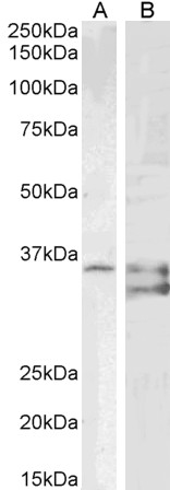
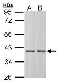
Figure 1. Western blot analysis of SGTA using anti-SGTA antibody (A03818-1). Electrophoresis was performed on a 5-20% SDS-PAGE gel at 70V (Stacking gel) / 90V (Resolving gel) for 2-3 hours. The sample well of each lane was loaded with 30 ug of sample under reducing conditions. Lane 1: human HepG2 whole cell lysates, Lane 2: human MOLT-4 whole cell lysates, Lane 3: human K562 whole cell lysates, Lane 4: human Hela whole cell lysates, Lane 5: rat liver tissue lysates, Lane 6: mouse liver tissue lysates. After electrophoresis, proteins were transferred to a nitrocellulose membrane at 150 mA for 50-90 minutes. Blocked the membrane with 5% non-fat milk/TBS for 1.5 hour at RT. The membrane was incubated with rabbit anti-SGTA antigen affinity purified polyclonal antibody (Catalog # A03818-1) at 0.25 microg/mL overnight at 4°C, then washed with TBS-0.1%Tween 3 times with 5 minutes each and probed with a goat anti-rabbit IgG-HRP secondary antibody at a dilution of 1:5000 for 1.5 hour at RT. The signal is developed using an Enhanced Chemiluminescent detection (ECL) kit (Catalog # EK1002) with Tanon 5200 system. A specific band was detected for SGTA at approximately 37 kDa. The expected band size for SGTA is at 34 kDa.
 and anti-Tubulin alpha antibody (M03989-3). SGTA and Tubulin alpha were detected in immunocytochemical section of U2OS cells. Enzyme antigen retrieval was performed using IHC enzyme antigen retrieval reagent (AR0022) for 15 mins. The cells were blocked with 10% goat serum. And then incubated with 5microg/mL rabbit anti-SGTA antibody (A03818-1) and mouse anti-Tubulin alpha Antibody (M03989-3) overnight at 4°C. Cy3 Conjugated Goat Anti-Rabbit IgG (BA1032) and DyLight®488 Conjugated Goat Anti-Mouse IgG (BA1126) were used as secondary antibody at 1:500 dilution and incubated for 30 minutes at 37°C. Visualize using a fluorescence microscope and filter sets appropriate for the label used.")
. Overlay histogram showing Jurkat cells stained with A03818-1 (Blue line). To facilitate intracellular staining, cells were fixed with 4% paraformaldehyde and permeabilized with permeabilization buffer. The cells were blocked with 10% normal goat serum. And then incubated with rabbit anti-SGTA Antibody (A03818-1, 1 microg/1x106 cells) for 30 min at 20°C. DyLight®488 conjugated goat anti-rabbit IgG (BA1127, 5-10 microg/1x106 cells) was used as secondary antibody for 30 minutes at 20°C. Isotype control antibody (Green line) was rabbit IgG (1 microg/1x106) used under the same conditions. Unlabelled sample (Red line) was also used as a control.")
Figure 1. Western blot analysis of SGTA using anti-SGTA antibody (A03818-1). Electrophoresis was performed on a 5-20% SDS-PAGE gel at 70V (Stacking gel) / 90V (Resolving gel) for 2-3 hours. The sample well of each lane was loaded with 30 ug of sample under reducing conditions. Lane 1: human HepG2 whole cell lysates, Lane 2: human MOLT-4 whole cell lysates, Lane 3: human K562 whole cell lysates, Lane 4: human Hela whole cell lysates, Lane 5: rat liver tissue lysates, Lane 6: mouse liver tissue lysates. After electrophoresis, proteins were transferred to a nitrocellulose membrane at 150 mA for 50-90 minutes. Blocked the membrane with 5% non-fat milk/TBS for 1.5 hour at RT. The membrane was incubated with rabbit anti-SGTA antigen affinity purified polyclonal antibody (Catalog # A03818-1) at 0.25 microg/mL overnight at 4°C, then washed with TBS-0.1%Tween 3 times with 5 minutes each and probed with a goat anti-rabbit IgG-HRP secondary antibody at a dilution of 1:5000 for 1.5 hour at RT. The signal is developed using an Enhanced Chemiluminescent detection (ECL) kit (Catalog # EK1002) with Tanon 5200 system. A specific band was detected for SGTA at approximately 37 kDa. The expected band size for SGTA is at 34 kDa.
Anti-SGTA Antibody Picoband(r)
A03818-1-CARRIER-FREE
ApplicationsFlow Cytometry, ImmunoFluorescence, Western Blot, ELISA, ImmunoCytoChemistry
Product group Antibodies
ReactivityHuman, Mouse, Rat
TargetSGTA
Overview
- SupplierBoster Bio
- Product NameAnti-SGTA Antibody Picoband(r)
- Delivery Days Customer9
- ApplicationsFlow Cytometry, ImmunoFluorescence, Western Blot, ELISA, ImmunoCytoChemistry
- CertificationResearch Use Only
- ClonalityPolyclonal
- Concentration500 ug/ml
- Gene ID6449
- Target nameSGTA
- Target descriptionsmall glutamine rich tetratricopeptide repeat co-chaperone alpha
- Target synonymsSGT, SGT1, Vpu, alphaSGT, hSGT, small glutamine-rich tetratricopeptide repeat-containing protein alpha, UBP, alpha-SGT, protein containing three tetratricopeptide repeats, small glutamine rich tetratricopeptide repeat containing alpha, small glutamine-rich tetratricopeptide repeat (TPR)-containing, alpha, viral protein U-binding protein, vpu-binding protein
- HostRabbit
- IsotypeIgG
- Protein IDO43765
- Protein NameSmall glutamine-rich tetratricopeptide repeat-containing protein alpha
- Scientific DescriptionBoster Bio Anti-SGTA Antibody Picoband® catalog # A03818-1. Tested in ELISA, Flow Cytometry, IF, ICC, WB applications. This antibody reacts with Human, Mouse, Rat. The brand Picoband indicates this is a premium antibody that guarantees superior quality, high affinity, and strong signals with minimal background in Western blot applications. Only our best-performing antibodies are designated as Picoband, ensuring unmatched performance.
- ReactivityHuman, Mouse, Rat
- Storage Instruction-20°C,2°C to 8°C
- UNSPSC12352203
Related products
Product group Antibodies
SGTA AntibodyCSB-PA021204ESR2HU
ApplicationsImmunoFluorescence, ELISA, ImmunoHistoChemistry
ReactivityHuman
TargetSGTA
- SizePrice
Product group Antibodies
Anti-SGTA AntibodyA85083
ApplicationsWestern Blot, ELISA, ImmunoHistoChemistry
ReactivityHuman, Mouse
- SizePrice
Product group Antibodies

SGTA / SGT AntibodyLS-C830980
ApplicationsELISA, ImmunoHistoChemistry
ReactivityHuman, Mouse, Rat
TargetSGTA
- SizePrice

Product group Antibodies
Anti-SGTA AntibodyHPA056309
ApplicationsWestern Blot, ImmunoCytoChemistry, ImmunoHistoChemistry
ReactivityHuman
TargetSGTA
- SizePrice
Product group Antibodies

ApplicationsWestern Blot, ELISA, ImmunoHistoChemistry
ReactivityBovine, Canine, Human, Mouse, Porcine, Rat
TargetSGTA
- SizePrice
Product group Antibodies
SGTA antibodyGTX101532
ApplicationsWestern Blot, ImmunoHistoChemistry, ImmunoHistoChemistry Paraffin
ReactivityHuman, Mouse
TargetSGTA
- SizePrice
Product group Antibodies
Anti-SGTA Antibody144-07306
ApplicationsImmunoFluorescence, Western Blot, ImmunoHistoChemistry
ReactivityHuman
TargetSGTA
- SizePrice
Product group Antibodies
SGTA Polyclonal AntibodyBS-55197R
ApplicationsImmunoFluorescence, Western Blot, ImmunoCytoChemistry, ImmunoHistoChemistry, ImmunoHistoChemistry Paraffin
ReactivityHuman
TargetSGTA
- SizePrice