


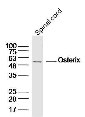
Figure 1. Western blot analysis of Sp7/Osterix using anti-Sp7/Osterix antibody (A02077-1). Electrophoresis was performed on a 5-20% SDS-PAGE gel at 70V (Stacking gel) / 90V (Resolving gel) for 2-3 hours. The sample well of each lane was loaded with 50ug of sample under reducing conditions. Lane 1: human HELA whole cell lysates, Lane 2: human U87 whole cell lysates, Lane 3: human MDA-MB453 whole cell lysates, Lane 4: human MCF-7 whole cell lysates, Lane 5: human Jurkat whole cell lysates, Lane 6: human THP-1 whole cell lysates. Lane 7: rat lung tissue lysates, Lane 8: rat thymus tissue lysates, Lane 9: rat skeletal muscle tissue lysates, Lane 10: rat testis tissue lysates, Lane 11: mouse lung tissue lysates, Lane 12: mouse thymus tissue lysates. After Electrophoresis, proteins were transferred to a Nitrocellulose membrane at 150mA for 50-90 minutes. Blocked the membrane with 5% Non-fat Milk/ TBS for 1.5 hour at RT. The membrane was incubated with rabbit anti-Sp7/Osterix antigen affinity purified polyclonal antibody (Catalog # A02077-1) at 0.5 microg/mL overnight at 4°C, then washed with TBS-0.1%Tween 3 times with 5 minutes each and probed with a goat anti-rabbit IgG-HRP secondary antibody at a dilution of 1:5000 for 1.5 hour at RT. The signal is developed using an Enhanced Chemiluminescent detection (ECL) kit (Catalog # EK1002) with Tanon 5200 system. A specific band was detected for Sp7/Osterix at approximately 45KD. The expected band size for Sp7/Osterix is at 45KD.
Figure 1. Western blot analysis of Sp7/Osterix using anti-Sp7/Osterix antibody (A02077-1). Electrophoresis was performed on a 5-20% SDS-PAGE gel at 70V (Stacking gel) / 90V (Resolving gel) for 2-3 hours. The sample well of each lane was loaded with 50ug of sample under reducing conditions. Lane 1: human HELA whole cell lysates, Lane 2: human U87 whole cell lysates, Lane 3: human MDA-MB453 whole cell lysates, Lane 4: human MCF-7 whole cell lysates, Lane 5: human Jurkat whole cell lysates, Lane 6: human THP-1 whole cell lysates. Lane 7: rat lung tissue lysates, Lane 8: rat thymus tissue lysates, Lane 9: rat skeletal muscle tissue lysates, Lane 10: rat testis tissue lysates, Lane 11: mouse lung tissue lysates, Lane 12: mouse thymus tissue lysates. After Electrophoresis, proteins were transferred to a Nitrocellulose membrane at 150mA for 50-90 minutes. Blocked the membrane with 5% Non-fat Milk/ TBS for 1.5 hour at RT. The membrane was incubated with rabbit anti-Sp7/Osterix antigen affinity purified polyclonal antibody (Catalog # A02077-1) at 0.5 microg/mL overnight at 4°C, then washed with TBS-0.1%Tween 3 times with 5 minutes each and probed with a goat anti-rabbit IgG-HRP secondary antibody at a dilution of 1:5000 for 1.5 hour at RT. The signal is developed using an Enhanced Chemiluminescent detection (ECL) kit (Catalog # EK1002) with Tanon 5200 system. A specific band was detected for Sp7/Osterix at approximately 45KD. The expected band size for Sp7/Osterix is at 45KD.
Anti-Sp7/Osterix Antibody Picoband(r)
A02077-1-BIOTIN
ApplicationsWestern Blot, ELISA
Product group Antibodies
ReactivityHuman, Mouse, Rat
TargetSP7
Overview
- SupplierBoster Bio
- Product NameAnti-Sp7/Osterix Antibody Picoband(r)
- Delivery Days Customer9
- ApplicationsWestern Blot, ELISA
- CertificationResearch Use Only
- ClonalityPolyclonal
- Concentration500 ug/ml
- ConjugateBiotin
- Gene ID121340
- Target nameSP7
- Target descriptionSp7 transcription factor
- Target synonymsOI11, OI12, OSX, osterix, transcription factor Sp7, zinc finger protein osterix
- HostRabbit
- IsotypeIgG
- Protein IDQ8TDD2
- Protein NameTranscription factor Sp7
- Scientific DescriptionBoster Bio Anti-Sp7/Osterix Antibody Picoband® catalog # A02077-1. Tested in ELISA, WB applications. This antibody reacts with Human, Mouse, Rat. The brand Picoband indicates this is a premium antibody that guarantees superior quality, high affinity, and strong signals with minimal background in Western blot applications. Only our best-performing antibodies are designated as Picoband, ensuring unmatched performance.
- ReactivityHuman, Mouse, Rat
- Storage Instruction-20°C,2°C to 8°C
- UNSPSC12352203
Related products
Product group Antibodies
ApplicationsImmunoPrecipitation, Western Blot, ImmunoCytoChemistry, ImmunoHistoChemistry
TargetSP7
- SizePrice
Product group Antibodies
References
SP7/Osterix Polyclonal AntibodyBS-1110R
ApplicationsWestern Blot, ELISA, ImmunoHistoChemistry, ImmunoHistoChemistry Paraffin
TargetSP7
- SizePrice
Product group Antibodies
Anti-SP7 Antibody101-11645
ApplicationsImmunoFluorescence, Western Blot, ELISA
TargetSP7
- SizePrice
Product group Antibodies

Anti-SP7 [RAB-S202]Ab01890-1.1
ApplicationsImmunoPrecipitation
ReactivityHuman
TargetSP7
- SizePrice
Product group Antibodies
SP7 antibodyGTX129385
ApplicationsWestern Blot
ReactivityHuman
TargetSP7
- SizePrice
Product group Antibodies

SP7 / Osterix AntibodyLS-C780018
ApplicationsWestern Blot, ELISA
ReactivityHuman, Mouse
TargetSP7
- SizePrice
Product group Antibodies
Anti-SP7 AntibodyHPA063202
ApplicationsImmunoHistoChemistry
ReactivityHuman
TargetSP7
- SizePrice
Product group Antibodies
Anti-Sp7/Osterix Antibody Picoband(r)A02077-1-CARRIER-FREE
ApplicationsWestern Blot, ELISA
ReactivityHuman, Mouse, Rat
TargetSP7
- SizePrice