

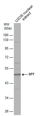
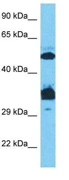
U2OS nuclear extract (30 μg) was separated by 10% SDS-PAGE, and the membrane was blotted with SP7 antibody (GTX129385) diluted at 1:500.
U2OS nuclear extract (30 μg) was separated by 10% SDS-PAGE, and the membrane was blotted with SP7 antibody (GTX129385) diluted at 1:500.
SP7 antibody
GTX129385
ApplicationsWestern Blot
Product group Antibodies
ReactivityHuman
TargetSP7
Overview
- SupplierGeneTex
- Product NameSP7 antibody
- Delivery Days Customer9
- Application Supplier NoteWB: 1:500-1:3000. *Optimal dilutions/concentrations should be determined by the researcher.Not tested in other applications.
- ApplicationsWestern Blot
- CertificationResearch Use Only
- ClonalityPolyclonal
- Concentration1 mg/ml
- ConjugateUnconjugated
- Gene ID121340
- Target nameSP7
- Target descriptionSp7 transcription factor
- Target synonymsOI11, OI12, OSX, osterix, transcription factor Sp7, zinc finger protein osterix
- HostRabbit
- IsotypeIgG
- Protein IDQ8TDD2
- Protein NameTranscription factor Sp7
- Scientific DescriptionSP7 is a C2H2-type zinc finger transcription factor of the SP gene family and a putative master regulator of bone cell differentiation (Gao et al., 2004 [PubMed 15474293]).[supplied by OMIM]
- ReactivityHuman
- Storage Instruction-20°C or -80°C,2°C to 8°C
- UNSPSC41116161
Datasheet
Related products
Product group Antibodies

Anti-SP7 [RAB-S202]Ab01890-1.1
ApplicationsImmunoPrecipitation
ReactivityHuman
TargetSP7
- SizePrice
Product group Antibodies
Anti-SP7 Antibody101-11645
ApplicationsImmunoFluorescence, Western Blot, ELISA
TargetSP7
- SizePrice

Product group Antibodies
Anti-Sp7/Osterix Antibody Picoband(r)A02077-1-CARRIER-FREE
ApplicationsWestern Blot, ELISA
ReactivityHuman, Mouse, Rat
TargetSP7
- SizePrice

Product group Antibodies
References
SP7/Osterix Polyclonal AntibodyBS-1110R
ApplicationsWestern Blot, ELISA, ImmunoHistoChemistry, ImmunoHistoChemistry Paraffin
TargetSP7
- SizePrice
Product group Antibodies
ApplicationsImmunoPrecipitation, Western Blot, ImmunoCytoChemistry, ImmunoHistoChemistry
TargetSP7
- SizePrice
Product group Antibodies
SP7 antibodyGTX02884
ApplicationsWestern Blot, ImmunoHistoChemistry, ImmunoHistoChemistry Paraffin
ReactivityHuman, Mouse, Rabbit
TargetSP7
- SizePrice
Product group Antibodies
SP7 antibodyGTX04567
ApplicationsWestern Blot
ReactivityHuman, Mouse
TargetSP7
- SizePrice
Product group Antibodies
Anti-SP7 AntibodyHPA063202
ApplicationsImmunoHistoChemistry
ReactivityHuman
TargetSP7
- SizePrice
Product group Antibodies
References
SP7 antibody, C-termGTX50962
ApplicationsImmunoFluorescence, Western Blot, ImmunoCytoChemistry, ImmunoHistoChemistry, ImmunoHistoChemistry Paraffin
ReactivityHuman
TargetSP7
- SizePrice
Product group Antibodies
SP7 antibodyGTX37579
ApplicationsImmunoHistoChemistry, ImmunoHistoChemistry Paraffin
ReactivityHuman, Mouse
TargetSP7
- SizePrice