


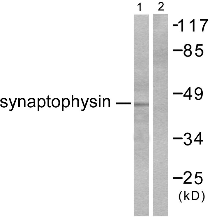
Figure 1. Western blot analysis of SYP using anti-SYP antibody (M05049-3). Electrophoresis was performed on a 5-20% SDS-PAGE gel at 70V (Stacking gel) / 90V (Resolving gel) for 2-3 hours. The sample well of each lane was loaded with 50ug of sample under reducing conditions. Lane 1: rat brain tissue lysates, Lane 2: rat brain whole cell lysates, Lane 3: mouse brain whole cell lysates, Lane 4: mouse brain whole cell lysates, After Electrophoresis, proteins were transferred to a Nitrocellulose membrane at 150mA for 50-90 minutes. Blocked the membrane with 5% Non-fat Milk/ TBS for 1.5 hour at RT. The membrane was incubated with mouse anti-SYP antigen affinity purified polyclonal antibody (Catalog # M05049-3) at 0.5 g/mL overnight at 4C, then washed with TBS-0.1%Tween 3 times with 5 minutes each and probed with a goat anti-mouse IgG-HRP secondary antibody at a dilution of 1:10000 for 1.5 hour at RT. The signal is developed using an Enhanced Chemiluminescent detection (ECL) kit (Catalog # EK1001) with Tanon 5200 system. A specific band was detected for SYP at approximately 38KD. The expected band size for SYP is at 34KD.
Figure 1. Western blot analysis of SYP using anti-SYP antibody (M05049-3). Electrophoresis was performed on a 5-20% SDS-PAGE gel at 70V (Stacking gel) / 90V (Resolving gel) for 2-3 hours. The sample well of each lane was loaded with 50ug of sample under reducing conditions. Lane 1: rat brain tissue lysates, Lane 2: rat brain whole cell lysates, Lane 3: mouse brain whole cell lysates, Lane 4: mouse brain whole cell lysates, After Electrophoresis, proteins were transferred to a Nitrocellulose membrane at 150mA for 50-90 minutes. Blocked the membrane with 5% Non-fat Milk/ TBS for 1.5 hour at RT. The membrane was incubated with mouse anti-SYP antigen affinity purified polyclonal antibody (Catalog # M05049-3) at 0.5 g/mL overnight at 4C, then washed with TBS-0.1%Tween 3 times with 5 minutes each and probed with a goat anti-mouse IgG-HRP secondary antibody at a dilution of 1:10000 for 1.5 hour at RT. The signal is developed using an Enhanced Chemiluminescent detection (ECL) kit (Catalog # EK1001) with Tanon 5200 system. A specific band was detected for SYP at approximately 38KD. The expected band size for SYP is at 34KD.
Anti-SYP/Synaptophysin Antibody Picoband(r) (monoclonal, 3G12)
M05049-3-BIOTIN
ApplicationsWestern Blot
Product group Antibodies
ReactivityMouse, Rat
TargetSYP
Overview
- SupplierBoster Bio
- Product NameAnti-SYP/Synaptophysin Antibody Picoband(r) (monoclonal, 3G12)
- Delivery Days Customer9
- ApplicationsWestern Blot
- CertificationResearch Use Only
- ClonalityMonoclonal
- Clone ID3G12
- Concentration500 ug/ml
- ConjugateBiotin
- Gene ID6855
- Target nameSYP
- Target descriptionsynaptophysin
- Target synonymsMRX96, MRXSYP, XLID96, synaptophysin, major synaptic vesicle protein P38
- HostMouse
- IsotypeIgG2b
- Protein IDP07825
- Protein NameSynaptophysin
- Scientific DescriptionBoster Bio Anti-SYP/Synaptophysin Antibody Picoband® (monoclonal, 3G12) catalog # M05049-3. Tested in WB applications. This antibody reacts with Mouse, Rat. The brand Picoband indicates this is a premium antibody that guarantees superior quality, high affinity, and strong signals with minimal background in Western blot applications. Only our best-performing antibodies are designated as Picoband, ensuring unmatched performance.
- ReactivityMouse, Rat
- Storage Instruction-20°C,2°C to 8°C
- UNSPSC12352203
Related products
Product group Antibodies
ApplicationsImmunoPrecipitation, Western Blot, ImmunoCytoChemistry, ImmunoHistoChemistry
TargetSYP
- SizePrice
Product group Antibodies
Anti-SYP Antibody144-06344
ApplicationsImmunoFluorescence, Western Blot, ImmunoHistoChemistry
ReactivityHuman, Mouse, Rat
TargetSYP
- SizePrice
Product group Antibodies
References
Synaptophysin antibodyGTX100865
ApplicationsImmunoFluorescence, Western Blot, ImmunoCytoChemistry, ImmunoHistoChemistry, ImmunoHistoChemistry Frozen, ImmunoHistoChemistry Paraffin
ReactivityHuman, Mouse, Rat
TargetSYP
- SizePrice
Product group Antibodies

Synaptophysin (SYP) AntibodyABX013199
ApplicationsWestern Blot, ELISA, ImmunoHistoChemistry
- SizePrice
Product group Antibodies
ApplicationsWestern Blot, ELISA, ImmunoHistoChemistry
ReactivityHuman, Mouse, Rat
- SizePrice

Product group Antibodies
Anti-Synaptophysin/SYP Picoband(r) AntibodyA05049-CARRIER-FREE
ApplicationsImmunoFluorescence, Western Blot, ImmunoCytoChemistry, ImmunoHistoChemistry
ReactivityHuman, Mouse, Rat
TargetSYP
- SizePrice
Product group Antibodies
SYP AntibodyCSB-PA004215
ApplicationsWestern Blot, ELISA, ImmunoHistoChemistry
ReactivityHuman, Mouse, Rat
TargetSYP
- SizePrice
Product group Antibodies
anti-Synaptophysin (human), Rabbit Monoclonal (RM258)REV-31-1139-00
ApplicationsWestern Blot, ImmunoHistoChemistry
ReactivityHuman
TargetSYP
- SizePrice
Product group Antibodies

ApplicationsImmunoHistoChemistry
ReactivityHuman
TargetSYP
- SizePrice

Product group Antibodies
Synaptophysin Recombinant AntibodyBSM-52379R
ApplicationsFlow Cytometry, ImmunoFluorescence, Western Blot, ImmunoCytoChemistry, ImmunoHistoChemistry, ImmunoHistoChemistry Frozen, ImmunoHistoChemistry Paraffin
ReactivityHuman, Mouse, Rat
TargetSYP
- SizePrice