


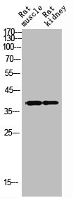
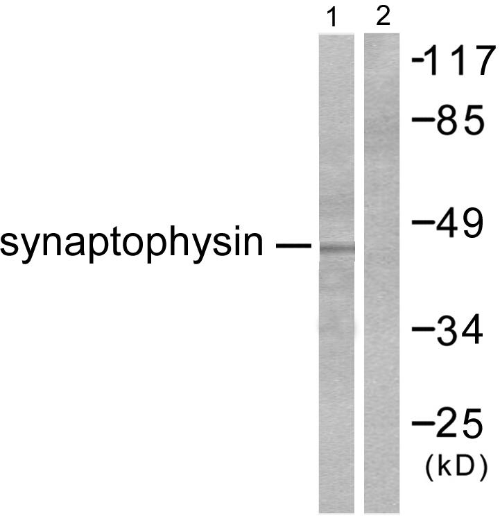
Lane 1:Human Cerebrum lysates; Lane 2: PC-12 cell lysates probed with Synaptophysin (2B1) Monoclonal Antibody (bsm-52379R) at 1:1000 dilution and 4˚C overnight incubation. Followed by conjugated secondary antibody incubation for 60 min at 37˚C.
 staining with Synaptophysin (2B1) Monoclonal Antibody (bsm-52379R) at 1:100 in N2A cells (red). The nuclear counterstain is DAPI (blue). Cells were fixed in paraformaldehyde, permeabilized with 0.25% Triton X100/PBS.")
 staining with Synaptophysin (2B1) Monoclonal Antibody (bsm-52379R) at 1:100 in PC-12 cells (red). The nuclear counterstain is DAPI (blue). Cells were fixed in paraformaldehyde, permeabilized with 0.25% Triton X100/PBS.")
 Monoclonal Antibody (bsm-52379R) at 1:100, overnight at 4°C, followed by a conjugated secondary antibody and DAB staining. Counterstained with hematoxylin.")
 Monoclonal Antibody (bsm-52379R) at 1:100, overnight at 4°C, followed by a conjugated secondary antibody and DAB staining. Counterstained with hematoxylin.")
 Monoclonal Antibody (bsm-52379R) at 1:100, overnight at 4°C, followed by a conjugated secondary antibody and DAB staining. Counterstained with hematoxylin.")
 for 15min; Block endogenous peroxidase by 3% hydrogen peroxide for 20 minutes; Blocking buffer (normal goat serum) at 37°C for 30min; Antibody incubation with Synaptophysin (2B1) Monoclonal Antibody, Unconjugated (bsm-52379R) at 1:200 overnight at 4°C, DAB staining.")
 for 15min; Block endogenous peroxidase by 3% hydrogen peroxide for 20 minutes; Blocking buffer (normal goat serum) at 37°C for 30min; Antibody incubation with Synaptophysin (2B1) Monoclonal Antibody, Unconjugated (bsm-52379R) at 1:200 overnight at 4°C, DAB staining.")
 for 15min; Block endogenous peroxidase by 3% hydrogen peroxide for 20 minutes; Blocking buffer (normal goat serum) at 37°C for 30min; Antibody incubation with Synaptophysin (2B1) Monoclonal Antibody, Unconjugated (bsm-52379R) at 1:200 overnight at 4°C, DAB staining.")
 for 15min; Block endogenous peroxidase by 3% hydrogen peroxide for 20 minutes; Blocking buffer (normal goat serum) at 37°C for 30min; Antibody incubation with Synaptophysin (2B1) Monoclonal Antibody, Unconjugated (bsm-52379R) at 1:200 overnight at 4°C, DAB staining.")
Lane 1:Human Cerebrum lysates; Lane 2: PC-12 cell lysates probed with Synaptophysin (2B1) Monoclonal Antibody (bsm-52379R) at 1:1000 dilution and 4˚C overnight incubation. Followed by conjugated secondary antibody incubation for 60 min at 37˚C.
Synaptophysin Recombinant Antibody
BSM-52379R
ApplicationsFlow Cytometry, ImmunoFluorescence, Western Blot, ImmunoHistoChemistry, ImmunoHistoChemistry Frozen, ImmunoHistoChemistry Paraffin
Product group Antibodies
ReactivityHuman, Mouse, Rat
TargetSYP
Overview
- SupplierBioss
- Product NameSynaptophysin Recombinant Antibody
- Delivery Days Customer16
- ApplicationsFlow Cytometry, ImmunoFluorescence, Western Blot, ImmunoHistoChemistry, ImmunoHistoChemistry Frozen, ImmunoHistoChemistry Paraffin
- Applications SupplierWB(1:300-5000), IHC-P(1:200-400), IHC-F(1:100-500), IF(), FCM(1ug/Test)
- CertificationResearch Use Only
- ClonalityMonoclonal
- Concentration1 mg/ml
- ConjugateUnconjugated
- Gene ID6855
- Target nameSYP
- Target descriptionsynaptophysin
- Target synonymsMRX96, MRXSYP, XLID96, synaptophysin, major synaptic vesicle protein P38
- HostRabbit
- IsotypeIgG
- Protein IDP08247
- Protein NameSynaptophysin
- ReactivityHuman, Mouse, Rat
- Storage Instruction-20°C
- UNSPSC41116161
Datasheet
Related products
Product group Antibodies
SYP AntibodyCSB-PA004215
ApplicationsWestern Blot, ELISA, ImmunoHistoChemistry
ReactivityHuman, Mouse, Rat
TargetSYP
- SizePrice
Product group Antibodies
Anti-SYP Antibody144-06344
ApplicationsImmunoFluorescence, Western Blot, ImmunoHistoChemistry
ReactivityHuman, Mouse, Rat
TargetSYP
- SizePrice
Product group Antibodies
ApplicationsWestern Blot, ELISA, ImmunoHistoChemistry
ReactivityHuman, Mouse, Rat
- SizePrice
Product group Antibodies

Synaptophysin (SYP) AntibodyABX013199
ApplicationsWestern Blot, ELISA, ImmunoHistoChemistry
- SizePrice

Product group Antibodies
Anti-Synaptophysin/SYP Picoband(r) AntibodyA05049-CARRIER-FREE
ApplicationsImmunoFluorescence, Western Blot, ImmunoCytoChemistry, ImmunoHistoChemistry
ReactivityHuman, Mouse, Rat
TargetSYP
- SizePrice
Product group Antibodies

ApplicationsImmunoHistoChemistry
ReactivityHuman
TargetSYP
- SizePrice

Product group Antibodies
Anti-SYP AntibodyHPA002858
ApplicationsWestern Blot, ImmunoHistoChemistry
ReactivityHuman
TargetSYP
- SizePrice
![Synaptophysin antibody detects Synaptophysin protein at synaptic vesicles by immunofluorescent analysis. Sample: DIV9 rat E18 primary cortical neurons were fixed in 4% paraformaldehyde at RT for 15 min. Green: Synaptophysin protein stained by Synaptophysin antibody (GTX100865) diluted at 1:500. Red: beta Tubulin 3/ Tuj1, stained by beta Tubulin 3/ Tuj1 antibody [GT11710] (GTX631836) diluted at 1:500. Blue: Fluoroshield with DAPI (GTX30920).](https://www.genetex.com/upload/website/prouct_img/normal/GTX100865/GTX100865_40128_20170503_IFA_R_w_23060100_733.webp)
Product group Antibodies
Synaptophysin antibodyGTX100865
ApplicationsImmunoFluorescence, Western Blot, ImmunoCytoChemistry, ImmunoHistoChemistry, ImmunoHistoChemistry Frozen, ImmunoHistoChemistry Paraffin
ReactivityHuman, Mouse, Rat
TargetSYP
- SizePrice