


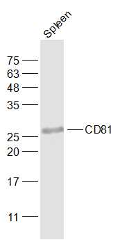
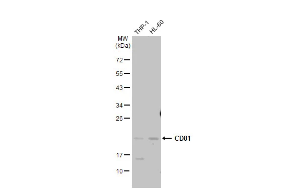
Figure 1. Western blot analysis of TAPA1 using anti-TAPA1 antibody (A01281-2). Electrophoresis was performed on a 5-20% SDS-PAGE gel at 70V (Stacking gel) / 90V (Resolving gel) for 2-3 hours. The sample well of each lane was loaded with 50ug of sample under reducing conditions. Lane 1: mouse raw264.7 whole cell lysates, After Electrophoresis, proteins were transferred to a Nitrocellulose membrane at 150mA for 50-90 minutes. Blocked the membrane with 5% Non-fat Milk/ TBS for 1.5 hour at RT. The membrane was incubated with rabbit anti-TAPA1 antigen affinity purified polyclonal antibody (Catalog # A01281-2) at 0.5 microg/mL overnight at 4°C, then washed with TBS-0.1%Tween 3 times with 5 minutes each and probed with a goat anti-rabbit IgG-HRP secondary antibody at a dilution of 1:10000 for 1.5 hour at RT. The signal is developed using an Enhanced Chemiluminescent detection (ECL) kit (Catalog # EK1002) with Tanon 5200 system. A specific band was detected for TAPA1 at approximately 22KD. The expected band size for TAPA1 is at 22KD.
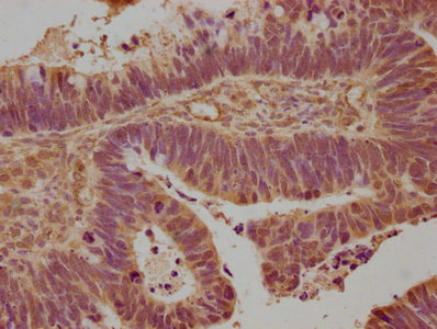
. TAPA1 was detected in paraffin-embedded section of human tonsil tissue . Heat mediated antigen retrieval was performed in citrate buffer (pH6, epitope retrieval solution) for 20 mins. The tissue section was blocked with 10% goat serum. The tissue section was then incubated with 1microg/ml rabbit anti-TAPA1 Antibody (A01281-2) overnight at 4°C. Biotinylated goat anti-rabbit IgG was used as secondary antibody and incubated for 30 minutes at 37°C. The tissue section was developed using Strepavidin-Biotin-Complex (SABC)(Catalog # SA1022) with DAB as the chromogen.")
. Overlay histogram showing HL-60 cells stained with A01281-2 (Blue line). The cells were fixed with 4% paraformaldehyde and blocked with 10% normal goat serum. And then incubated with rabbit anti-TAPA1 Antibody (A01281-2, 1 microg/1x106 cells) for 30 min at 20°C. DyLight®488 conjugated goat anti-rabbit IgG (BA1127, 5-10 microg/1x106 cells) was used as secondary antibody for 30 minutes at 20°C. Isotype control antibody (Green line) was rabbit IgG (1 microg/1x106) used under the same conditions. Unlabelled sample without incubation with primary antibody and secondary antibody (Red line) was used as a blank control.")
. Overlay histogram showing U2OS cells stained with A01281-2 (Blue line). The cells were fixed with 4% paraformaldehyde and blocked with 10% normal goat serum. And then incubated with rabbit anti-TAPA1 Antibody (A01281-2, 1 microg/1x106 cells) for 30 min at 20°C. DyLight®488 conjugated goat anti-rabbit IgG (BA1127, 5-10 microg/1x106 cells) was used as secondary antibody for 30 minutes at 20°C. Isotype control antibody (Green line) was rabbit IgG (1 microg/1x106) used under the same conditions. Unlabelled sample without incubation with primary antibody and secondary antibody (Red line) was used as a blank control.")
Figure 1. Western blot analysis of TAPA1 using anti-TAPA1 antibody (A01281-2). Electrophoresis was performed on a 5-20% SDS-PAGE gel at 70V (Stacking gel) / 90V (Resolving gel) for 2-3 hours. The sample well of each lane was loaded with 50ug of sample under reducing conditions. Lane 1: mouse raw264.7 whole cell lysates, After Electrophoresis, proteins were transferred to a Nitrocellulose membrane at 150mA for 50-90 minutes. Blocked the membrane with 5% Non-fat Milk/ TBS for 1.5 hour at RT. The membrane was incubated with rabbit anti-TAPA1 antigen affinity purified polyclonal antibody (Catalog # A01281-2) at 0.5 microg/mL overnight at 4°C, then washed with TBS-0.1%Tween 3 times with 5 minutes each and probed with a goat anti-rabbit IgG-HRP secondary antibody at a dilution of 1:10000 for 1.5 hour at RT. The signal is developed using an Enhanced Chemiluminescent detection (ECL) kit (Catalog # EK1002) with Tanon 5200 system. A specific band was detected for TAPA1 at approximately 22KD. The expected band size for TAPA1 is at 22KD.
Anti-TAPA1/CD81 Antibody Picoband(r)
A01281-2-FITC
ApplicationsFlow Cytometry, Western Blot, ELISA, ImmunoCytoChemistry, ImmunoHistoChemistry, ImmunoHistoChemistry Frozen
Product group Antibodies
ReactivityHuman, Mouse, Rat
TargetCD81
Overview
- SupplierBoster Bio
- Product NameAnti-TAPA1/CD81 Antibody Picoband(r)
- Delivery Days Customer9
- ApplicationsFlow Cytometry, Western Blot, ELISA, ImmunoCytoChemistry, ImmunoHistoChemistry, ImmunoHistoChemistry Frozen
- CertificationResearch Use Only
- ClonalityPolyclonal
- Concentration500 ug/ml
- ConjugateFITC
- Gene ID975
- Target nameCD81
- Target descriptionCD81 molecule
- Target synonymsCVID6, S5.7, TAPA1, TSPAN28, CD81 antigen, 26 kDa cell surface protein TAPA-1, CD81 antigen (target of antiproliferative antibody 1), tetraspanin-28, tspan-28
- HostRabbit
- IsotypeIgG
- Protein IDP60033
- Protein NameCD81 antigen
- Scientific DescriptionBoster Bio Anti-TAPA1/CD81 Antibody Picoband® catalog # A01281-2. Tested in ELISA, Flow Cytometry, IHC, IHC-F, ICC, WB applications. This antibody reacts with Human, Mouse, Rat. The brand Picoband indicates this is a premium antibody that guarantees superior quality, high affinity, and strong signals with minimal background in Western blot applications. Only our best-performing antibodies are designated as Picoband, ensuring unmatched performance.
- ReactivityHuman, Mouse, Rat
- Storage Instruction-20°C,2°C to 8°C
- UNSPSC12352203
Related products
Product group Antibodies
CD81 AntibodyCSB-PA004960NA01HU
ApplicationsELISA, ImmunoHistoChemistry
ReactivityHuman
TargetCD81
- SizePrice
Product group Antibodies
Anti-TAPA1/CD81 Antibody Picoband(r)A01281-2-CARRIER-FREE
ApplicationsFlow Cytometry, Western Blot, ELISA, ImmunoCytoChemistry, ImmunoHistoChemistry, ImmunoHistoChemistry Frozen
ReactivityHuman, Mouse, Rat
TargetCD81
- SizePrice
Product group Antibodies

Anti-CD81 [1D6]Ab00697-1.1
ApplicationsFunctional Assay, Flow Cytometry, ImmunoFluorescence, ImmunoPrecipitation, Western Blot, ELISA, ImmunoCytoChemistry, ImmunoHistoChemistry, ImmunoHistoChemistry Paraffin
ReactivityGoat, Human, Primate, Sheep
TargetCD81
- SizePrice

Product group Antibodies
Anti-CD81 AntibodyA98238
ApplicationsWestern Blot, ELISA
ReactivityHuman, Mouse, Rat
- SizePrice

Product group Antibodies
Anti-CD81 AntibodyHPA007234
ApplicationsImmunoCytoChemistry, ImmunoHistoChemistry
ReactivityHuman
TargetCD81
- SizePrice
Product group Antibodies

CD81 Antibody (PE)LS-C485188
ApplicationsFlow Cytometry
ReactivityHuman
TargetCD81
- SizePrice
Product group Antibodies
ApplicationsFlow Cytometry
TargetCD81
- SizePrice
Product group Antibodies
CD81 Polyclonal AntibodyBS-6934R
ApplicationsImmunoFluorescence, Western Blot, ELISA, ImmunoCytoChemistry, ImmunoHistoChemistry, ImmunoHistoChemistry Frozen, ImmunoHistoChemistry Paraffin
ReactivityHuman, Mouse, Rat
TargetCD81
- SizePrice
Product group Antibodies
CD81 antibodyGTX101766
ApplicationsFlow Cytometry, ImmunoFluorescence, Western Blot, ImmunoCytoChemistry, ImmunoHistoChemistry, ImmunoHistoChemistry Paraffin, Neutralisation/Blocking
ReactivityHuman, Mouse, Rat
TargetCD81
- SizePrice