

Anti-Tau [PC1C6]
Ab01120-2.0
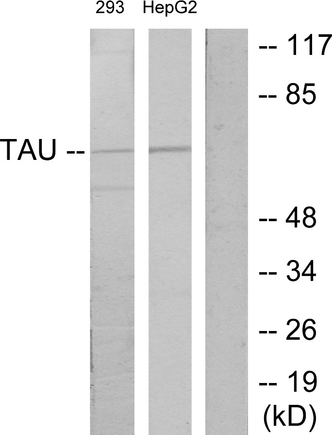
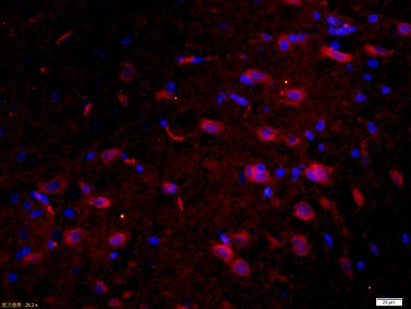
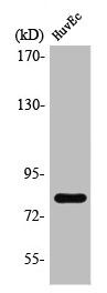
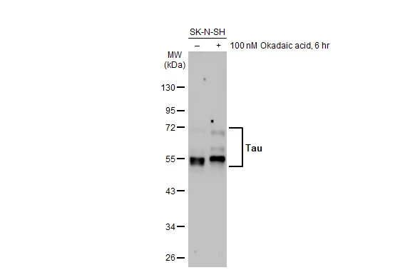
ApplicationsWestern Blot, ImmunoHistoChemistry
Product group Antibodies
ReactivityBovine, Human, Rat
TargetMAPT
Overview
- SupplierAbsolute Antibody
- Product NameAnti-Tau [PC1C6]
- Delivery Days Customer7
- Application Supplier NoteIn the original study, this antibody was used in immunoblot assays and competitive ELISA to show that tau was more abundant in bovine white matter extracts and microtubules than in extracts and microtubules from an enriched gray matter region of the brain (Binder et al., 1985). Example of functional studies in which this antibody could be utilised include those conducted by Kosik et al. (PNAS 1986) to confirm that tau is a major antigenic component of paired helical filaments in Alzheimer disease, by Bramblett et al. (Neuron 1993) to demonstrate that abnormal tau phosphorylation at Ser396 in Alzheimers disease recapitulates development and contributes to reduced microtubule binding, by Stamer et al. (J Cell Biol. 2002) to suggests a linkage between tau and amyloid precursor protein (APP) trafficking, and by Strang et al. (JBC 2018) to provide novel insights in the molecular mechanisms of tau aggregation.
- ApplicationsWestern Blot, ImmunoHistoChemistry
- Applications SupplierWB; IHC
- CertificationResearch Use Only
- ClonalityMonoclonal
- Clone IDPC1C6
- Gene ID4137
- Target nameMAPT
- Target descriptionmicrotubule associated protein tau
- Target synonymsDDPAC, FTD1, FTDP-17, MAPTL, MSTD, MTBT1, MTBT2, PPND, PPP1R103, TAU, Tau-PHF6, tau-40, microtubule-associated protein tau, G protein beta1/gamma2 subunit-interacting factor 1, PHF-tau, Tau-derived paired helical filament hexapeptide, neurofibrillary tangle protein, paired helical filament-tau, protein phosphatase 1, regulatory subunit 103
- HostMouse
- IsotypeIgG2a
- Protein IDP10636
- Protein NameMicrotubule-associated protein tau
- ReactivityBovine, Human, Rat
- Reactivity SupplierHuman, Rat, Bovine
- Reactivity Supplier NoteThis antibody was raised by immunising BALB/c mouse with purified denatured bovine microtubule associated proteins.
- Storage Instruction-20°C,2°C to 8°C
- UNSPSC41116161
Related products
Product group Antibodies
Anti-Tau AntibodyA101586
ApplicationsWestern Blot, ELISA
ReactivityHuman
- SizePrice
Product group Antibodies
Anti-Tau (213-222aa) Antibody130-10929
ApplicationsELISA
ReactivityHuman
TargetMAPT
- SizePrice

Product group Antibodies
Anti-Tau/MAPT Antibody Picoband(r)A00097-3-CARRIER-FREE
ApplicationsImmunoFluorescence, Western Blot, ELISA, ImmunoCytoChemistry
ReactivityHuman, Mouse, Rat
TargetMAPT
- SizePrice
Product group Antibodies
References
Tau Polyclonal AntibodyBS-0157R
ApplicationsFlow Cytometry, ImmunoFluorescence, Western Blot, ELISA, ImmunoCytoChemistry, ImmunoHistoChemistry, ImmunoHistoChemistry Frozen, ImmunoHistoChemistry Paraffin
ReactivityHuman, Mouse, Rabbit, Rat
TargetMAPT
- SizePrice
Product group Antibodies
MAPT AntibodyCSB-PA004230
ApplicationsWestern Blot, ELISA
ReactivityHuman, Mouse, Rat
TargetMAPT
- SizePrice
Product group Antibodies
Mapt Polyclonal AntibodyCAC07406
ApplicationsImmunoFluorescence, Western Blot, ELISA, ImmunoHistoChemistry
ReactivityMouse
TargetMAPT
- SizePrice
Product group Antibodies
Tau antibodyGTX100866
ApplicationsImmunoFluorescence, Western Blot, ImmunoCytoChemistry, ImmunoHistoChemistry, ImmunoHistoChemistry Paraffin
ReactivityHuman, Mouse, Rat
TargetMAPT
- SizePrice
Product group Antibodies

ApplicationsImmunoPrecipitation, Western Blot, ELISA
ReactivityHuman
TargetMAPT
- SizePrice

Product group Antibodies
Anti-MAPT AntibodyHPA069524
ApplicationsImmunoHistoChemistry
ReactivityHuman
TargetMAPT
- SizePrice