


Figure 1. Western blot analysis of Tau/MAPT using anti-Tau/MAPT antibody (A00097-3). Electrophoresis was performed on a 5-20% SDS-PAGE gel at 70V (Stacking gel) / 90V (Resolving gel) for 2-3 hours. The sample well of each lane was loaded with 30 ug of sample under reducing conditions. Lane 1: rat brain tissue lysates, Lane 2: mouse brain tissue lysates. After electrophoresis, proteins were transferred to a nitrocellulose membrane at 150 mA for 50-90 minutes. Blocked the membrane with 5% non-fat milk/TBS for 1.5 hour at RT. The membrane was incubated with rabbit anti-Tau/MAPT antigen affinity purified polyclonal antibody (Catalog # A00097-3) at 0.5 microg/mL overnight at 4°C, then washed with TBS-0.1%Tween 3 times with 5 minutes each and probed with a goat anti-rabbit IgG-HRP secondary antibody at a dilution of 1:5000 for 1.5 hour at RT. The signal is developed using an Enhanced Chemiluminescent detection (ECL) kit (Catalog # EK1002) with Tanon 5200 system. A specific band was detected for Tau/MAPT at approximately 50-70 kDa. The expected band size for Tau/MAPT is at 79 kDa.
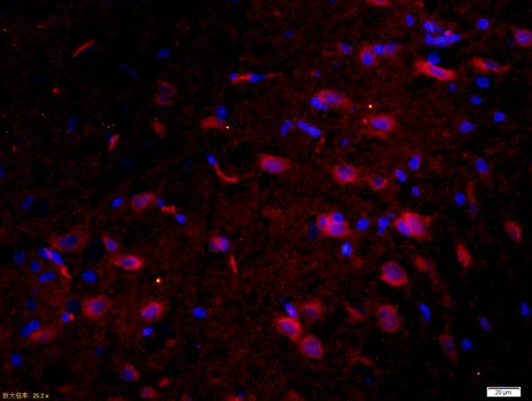
. Tau/MAPT was detected in an immunocytochemical section of T-47D cells. Enzyme antigen retrieval was performed using IHC enzyme antigen retrieval reagent (AR0022) for 15 mins. The cells were blocked with 10% goat serum. And then incubated with 5 microg/mL rabbit anti-Tau/MAPT Antibody (A00097-3) overnight at 4°C. DyLight®488 Conjugated Goat Anti-Rabbit IgG (BA1127) was used as secondary antibody at 1:100 dilution and incubated for 30 minutes at 37°C. The section was counterstained with DAPI. Visualize using a fluorescence microscope and filter sets appropriate for the label used.")
Figure 1. Western blot analysis of Tau/MAPT using anti-Tau/MAPT antibody (A00097-3). Electrophoresis was performed on a 5-20% SDS-PAGE gel at 70V (Stacking gel) / 90V (Resolving gel) for 2-3 hours. The sample well of each lane was loaded with 30 ug of sample under reducing conditions. Lane 1: rat brain tissue lysates, Lane 2: mouse brain tissue lysates. After electrophoresis, proteins were transferred to a nitrocellulose membrane at 150 mA for 50-90 minutes. Blocked the membrane with 5% non-fat milk/TBS for 1.5 hour at RT. The membrane was incubated with rabbit anti-Tau/MAPT antigen affinity purified polyclonal antibody (Catalog # A00097-3) at 0.5 microg/mL overnight at 4°C, then washed with TBS-0.1%Tween 3 times with 5 minutes each and probed with a goat anti-rabbit IgG-HRP secondary antibody at a dilution of 1:5000 for 1.5 hour at RT. The signal is developed using an Enhanced Chemiluminescent detection (ECL) kit (Catalog # EK1002) with Tanon 5200 system. A specific band was detected for Tau/MAPT at approximately 50-70 kDa. The expected band size for Tau/MAPT is at 79 kDa.
Anti-Tau/MAPT Antibody Picoband(r)
A00097-3-CARRIER-FREE
ApplicationsImmunoFluorescence, Western Blot, ELISA, ImmunoCytoChemistry
Product group Antibodies
ReactivityHuman, Mouse, Rat
TargetMAPT
Overview
- SupplierBoster Bio
- Product NameAnti-Tau/MAPT Antibody Picoband(r)
- Delivery Days Customer9
- ApplicationsImmunoFluorescence, Western Blot, ELISA, ImmunoCytoChemistry
- CertificationResearch Use Only
- ClonalityPolyclonal
- Concentration500 ug/ml
- Gene ID4137
- Target nameMAPT
- Target descriptionmicrotubule associated protein tau
- Target synonymsDDPAC, FTD1, FTDP-17, MAPTL, MSTD, MTBT1, MTBT2, PPND, PPP1R103, TAU, Tau-PHF6, tau-40, microtubule-associated protein tau, G protein beta1/gamma2 subunit-interacting factor 1, PHF-tau, Tau-derived paired helical filament hexapeptide, neurofibrillary tangle protein, paired helical filament-tau, protein phosphatase 1, regulatory subunit 103
- HostRabbit
- IsotypeIgG
- Protein IDP10636
- Protein NameMicrotubule-associated protein tau
- Scientific DescriptionBoster Bio Anti-Tau/MAPT Antibody Picoband® catalog # A00097-3. Tested in ELISA, IF, ICC, WB applications. This antibody reacts with Human, Mouse, Rat. The brand Picoband indicates this is a premium antibody that guarantees superior quality, high affinity, and strong signals with minimal background in Western blot applications. Only our best-performing antibodies are designated as Picoband, ensuring unmatched performance.
- ReactivityHuman, Mouse, Rat
- Storage Instruction-20°C,2°C to 8°C
- UNSPSC12352203
Related products
Product group Antibodies
Anti-Tau AntibodyA101586
ApplicationsWestern Blot, ELISA
ReactivityHuman
- SizePrice
Product group Antibodies
Anti-Tau (213-222aa) Antibody130-10929
ApplicationsELISA
ReactivityHuman
TargetMAPT
- SizePrice
Product group Antibodies

Anti-Tau [PC1C6]Ab01120-2.0
ApplicationsWestern Blot, ImmunoHistoChemistry
ReactivityBovine, Human, Rat
TargetMAPT
- SizePrice
Product group Antibodies
References
Tau Polyclonal AntibodyBS-0157R
ApplicationsFlow Cytometry, ImmunoFluorescence, Western Blot, ELISA, ImmunoCytoChemistry, ImmunoHistoChemistry, ImmunoHistoChemistry Frozen, ImmunoHistoChemistry Paraffin
ReactivityHuman, Mouse, Rabbit, Rat
TargetMAPT
- SizePrice
Product group Antibodies
MAPT AntibodyCSB-PA004230
ApplicationsWestern Blot, ELISA
ReactivityHuman, Mouse, Rat
TargetMAPT
- SizePrice
Product group Antibodies
Mapt Polyclonal AntibodyCAC07406
ApplicationsImmunoFluorescence, Western Blot, ELISA, ImmunoHistoChemistry
ReactivityMouse
TargetMAPT
- SizePrice
Product group Antibodies
Tau antibodyGTX100866
ApplicationsImmunoFluorescence, Western Blot, ImmunoCytoChemistry, ImmunoHistoChemistry, ImmunoHistoChemistry Paraffin
ReactivityHuman, Mouse, Rat
TargetMAPT
- SizePrice
Product group Antibodies

ApplicationsImmunoPrecipitation, Western Blot, ELISA
ReactivityHuman
TargetMAPT
- SizePrice

Product group Antibodies
Anti-MAPT AntibodyHPA069524
ApplicationsImmunoHistoChemistry
ReactivityHuman
TargetMAPT
- SizePrice