


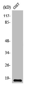
Figure 1. Western blot analysis of TGFA using anti-TGFA antibody (M01779-1). Electrophoresis was performed on a 5-20% SDS-PAGE gel at 70V (Stacking gel) / 90V (Resolving gel) for 2-3 hours. The sample well of each lane was loaded with 30 ug of sample under reducing conditions. Lane 1: human HepG2 whole cell lysates, Lane 2: human A431 whole cell lysates. After electrophoresis, proteins were transferred to a nitrocellulose membrane at 150 mA for 50-90 minutes. Blocked the membrane with 5% non-fat milk/TBS for 1.5 hour at RT. The membrane was incubated with rabbit anti-TGFA antigen affinity purified monoclonal antibody (Catalog # M01779-1) at 1:500 overnight at 4°C, then washed with TBS-0.1%Tween 3 times with 5 minutes each and probed with a goat anti-rabbit IgG-HRP secondary antibody at a dilution of 1:500 for 1.5 hour at RT. The signal is developed using an Enhanced Chemiluminescent detection (ECL) kit (Catalog # EK1002) with Tanon 5200 system. A specific band was detected for TGFA at approximately 17 kDa. The expected band size for TGFA is at 17 kDa.
Figure 1. Western blot analysis of TGFA using anti-TGFA antibody (M01779-1). Electrophoresis was performed on a 5-20% SDS-PAGE gel at 70V (Stacking gel) / 90V (Resolving gel) for 2-3 hours. The sample well of each lane was loaded with 30 ug of sample under reducing conditions. Lane 1: human HepG2 whole cell lysates, Lane 2: human A431 whole cell lysates. After electrophoresis, proteins were transferred to a nitrocellulose membrane at 150 mA for 50-90 minutes. Blocked the membrane with 5% non-fat milk/TBS for 1.5 hour at RT. The membrane was incubated with rabbit anti-TGFA antigen affinity purified monoclonal antibody (Catalog # M01779-1) at 1:500 overnight at 4°C, then washed with TBS-0.1%Tween 3 times with 5 minutes each and probed with a goat anti-rabbit IgG-HRP secondary antibody at a dilution of 1:500 for 1.5 hour at RT. The signal is developed using an Enhanced Chemiluminescent detection (ECL) kit (Catalog # EK1002) with Tanon 5200 system. A specific band was detected for TGFA at approximately 17 kDa. The expected band size for TGFA is at 17 kDa.
Anti-TGF alpha Monoclonal Antibody
M01779-1
ApplicationsImmunoFluorescence, ImmunoPrecipitation, Western Blot, ImmunoCytoChemistry, ImmunoHistoChemistry
Product group Antibodies
ReactivityHuman
TargetTGFA
Overview
- SupplierBoster Bio
- Product NameAnti-TGF alpha Monoclonal Antibody
- Delivery Days Customer9
- ApplicationsImmunoFluorescence, ImmunoPrecipitation, Western Blot, ImmunoCytoChemistry, ImmunoHistoChemistry
- CertificationResearch Use Only
- ClonalityMonoclonal
- Clone IDAEGF-20
- Gene ID7039
- Target nameTGFA
- Target descriptiontransforming growth factor alpha
- Target synonymsTFGA, protransforming growth factor alpha, TGF-alpha
- HostRabbit
- IsotypeIgG
- Protein IDP01135
- Protein NameProtransforming growth factor alpha
- Scientific DescriptionBoster Bio Anti-TGF alpha Monoclonal Antibody catalog # M01779-1. Tested in WB, IHC, ICC/IF, IP applications. This antibody reacts with Human.
- ReactivityHuman
- Storage Instruction-20°C
- UNSPSC12352203
References
- Liu F, Zhang J, Qin L, et al. Circular RNA EIF6 (Hsa_circ_0060060) sponges miR-144-3p to promote the cisplatin-resistance of human thyroid carcinoma cells by autophagy regulation. Aging (Albany NY). 2018,10(12):3806-3820. doi: 10.18632/aging.101674Read this paper
Datasheet
MSDS
Related products
Product group Antibodies
Anti-TGF alpha AntibodyA95170
ApplicationsWestern Blot, ELISA, ImmunoHistoChemistry
ReactivityHuman, Mouse, Rat
- SizePrice
Product group Antibodies

Anti-TGFalpha [tAb2]AB00614-2.0-BT
ApplicationsDot Blot, ELISA, Other Application
ReactivityHuman
TargetTGFA
- SizePrice
Product group Antibodies
Anti-TGF alpha Antibody188-10181
ReactivityBovine, Human, Mouse, Rat
TargetTGFA
- SizePrice
Product group Antibodies
References
TGF alpha Polyclonal AntibodyBS-0066R
ApplicationsWestern Blot, ELISA
ReactivityHuman, Mouse, Rat
TargetTGFA
- SizePrice
Product group Antibodies
TGFA AntibodyCSB-PA004275
ApplicationsWestern Blot, ELISA, ImmunoHistoChemistry
ReactivityHuman, Monkey, Mouse, Rat
TargetTGFA
- SizePrice
Product group Antibodies
TGFA Polyclonal AntibodyCAC14099
ApplicationsImmunoFluorescence, Western Blot, ELISA, ImmunoHistoChemistry
TargetTGFA
- SizePrice
Product group Antibodies

TGFA / TGF Alpha AntibodyLS-C401080
ApplicationsELISA, ImmunoHistoChemistry
ReactivityHuman, Mouse, Rat
TargetTGFA
- SizePrice

Product group Antibodies
Anti-TGFA AntibodyHPA042297
ApplicationsImmunoHistoChemistry
ReactivityHuman
TargetTGFA
- SizePrice
Product group Antibodies
TGF alpha antibodyGTX129103
ApplicationsWestern Blot, ImmunoHistoChemistry, ImmunoHistoChemistry Paraffin
ReactivityHuman
TargetTGFA
- SizePrice