


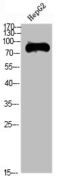
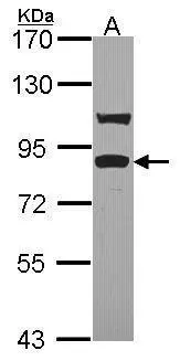
Figure 1. Western blot analysis of TRIF/TICAM1 using anti-TRIF/TICAM1 antibody (A01872-1). Electrophoresis was performed on a 5-20% SDS-PAGE gel at 70V (Stacking gel) / 90V (Resolving gel) for 2-3 hours. The sample well of each lane was loaded with 30ug of sample under reducing conditions. Lane 1: human Raji whole cell lysates. After Electrophoresis, proteins were transferred to a Nitrocellulose membrane at 150mA for 50-90 minutes. Blocked the membrane with 5% Non-fat Milk/ TBS for 1.5 hour at RT. The membrane was incubated with rabbit anti-TRIF/TICAM1 antigen affinity purified polyclonal antibody (Catalog # A01872-1) at 0.5 microg/mL overnight at 4°C, then washed with TBS-0.1%Tween 3 times with 5 minutes each and probed with a goat anti-rabbit IgG-HRP secondary antibody at a dilution of 1:5000 for 1.5 hour at RT. The signal is developed using an Enhanced Chemiluminescent detection (ECL) kit (Catalog # EK1002) with Tanon 5200 system. A specific band was detected for TRIF/TICAM1 at approximately 110KD. The expected band size for TRIF/TICAM1 is at 76KD.
. Overlay histogram showing PC-3 cells stained with A01872-1 (Blue line). To facilitate intracellular staining, cells were fixed with 4% paraformaldehyde and permeabilized with permeabilization buffer. The cells were blocked with 10% normal goat serum. And then incubated with rabbit anti-TRIF/TICAM1 Antibody (A01872-1, 1microg/1x106 cells) for 30 min at 20°C. DyLight®488 conjugated goat anti-rabbit IgG (BA1127, 5-10microg/1x106 cells) was used as secondary antibody for 30 minutes at 20°C. Isotype control antibody (Green line) was rabbit IgG (1microg/1x106) used under the same conditions. Unlabelled sample without incubation with primary antibody and secondary antibody (Red line) was used as a blank control.")
Figure 1. Western blot analysis of TRIF/TICAM1 using anti-TRIF/TICAM1 antibody (A01872-1). Electrophoresis was performed on a 5-20% SDS-PAGE gel at 70V (Stacking gel) / 90V (Resolving gel) for 2-3 hours. The sample well of each lane was loaded with 30ug of sample under reducing conditions. Lane 1: human Raji whole cell lysates. After Electrophoresis, proteins were transferred to a Nitrocellulose membrane at 150mA for 50-90 minutes. Blocked the membrane with 5% Non-fat Milk/ TBS for 1.5 hour at RT. The membrane was incubated with rabbit anti-TRIF/TICAM1 antigen affinity purified polyclonal antibody (Catalog # A01872-1) at 0.5 microg/mL overnight at 4°C, then washed with TBS-0.1%Tween 3 times with 5 minutes each and probed with a goat anti-rabbit IgG-HRP secondary antibody at a dilution of 1:5000 for 1.5 hour at RT. The signal is developed using an Enhanced Chemiluminescent detection (ECL) kit (Catalog # EK1002) with Tanon 5200 system. A specific band was detected for TRIF/TICAM1 at approximately 110KD. The expected band size for TRIF/TICAM1 is at 76KD.
Anti-TRIF/TICAM1 Antibody Picoband(r)
A01872-1-CARRIER-FREE
ApplicationsFlow Cytometry, Western Blot, ELISA
Product group Antibodies
ReactivityHuman
TargetTICAM1
Overview
- SupplierBoster Bio
- Product NameAnti-TRIF/TICAM1 Antibody Picoband(r)
- Delivery Days Customer9
- ApplicationsFlow Cytometry, Western Blot, ELISA
- CertificationResearch Use Only
- ClonalityPolyclonal
- Concentration500 ug/ml
- Gene ID148022
- Target nameTICAM1
- Target descriptionTIR domain containing adaptor molecule 1
- Target synonymsIIAE6, MyD88-3, PRVTIRB, TICAM-1, TRIF, TIR domain-containing adapter molecule 1, TIR domain containing adaptor inducing interferon-beta, TIR domain-containing adapter protein inducing IFN-beta, proline-rich, vinculin and TIR domain-containing protein B, putative NF-kappa-B-activating protein 502H, toll like receptor adaptor molecule 1, toll-interleukin-1 receptor domain-containing adapter protein inducing interferon beta
- HostRabbit
- IsotypeIgG
- Protein IDQ8IUC6
- Protein NameTIR domain-containing adapter molecule 1
- Scientific DescriptionBoster Bio Anti-TRIF/TICAM1 Antibody Picoband® catalog # A01872-1. Tested in ELISA, Flow Cytometry, WB applications. This antibody reacts with Human. The brand Picoband indicates this is a premium antibody that guarantees superior quality, high affinity, and strong signals with minimal background in Western blot applications. Only our best-performing antibodies are designated as Picoband, ensuring unmatched performance.
- ReactivityHuman
- Storage Instruction-20°C,2°C to 8°C
- UNSPSC12352203
Related products
Product group Antibodies
Anti-TRIF AntibodyA286020
ApplicationsFlow Cytometry, ELISA, ImmunoHistoChemistry
ReactivityHuman
- SizePrice
Product group Antibodies
Anti-TICAM1 Antibody144-60070
ApplicationsWestern Blot
ReactivityHuman, Mouse, Rat
TargetTICAM1
- SizePrice
Product group Antibodies

TICAM1 / TRIF AntibodyLS-C748647
ApplicationsWestern Blot, ImmunoHistoChemistry
ReactivityHuman, Mouse, Rat
TargetTICAM1
- SizePrice
Product group Antibodies

Goat anti-TICAM1EB09579
ApplicationsFlow Cytometry, ELISA, ImmunoHistoChemistry
ReactivityHuman
TargetTICAM1
- SizePrice
Product group Antibodies
TICAM1 AntibodyCSB-PA544544
ApplicationsWestern Blot, ELISA
ReactivityHuman
TargetTICAM1
- SizePrice
Product group Antibodies
ApplicationsImmunoPrecipitation, Western Blot, ImmunoCytoChemistry, ImmunoHistoChemistry
ReactivityMouse
TargetTICAM1
- SizePrice

Product group Antibodies
Anti-TICAM1 AntibodyHPA042460
ApplicationsImmunoHistoChemistry
ReactivityHuman
TargetTICAM1
- SizePrice
Product group Antibodies
TRIF antibody [C3], C-termGTX104744
ApplicationsImmunoFluorescence, Western Blot, ImmunoCytoChemistry
ReactivityHuman
TargetTICAM1
- SizePrice
Product group Antibodies
Anti-TICAM1 AntibodyCAB13605
ApplicationsWestern Blot, ELISA
ReactivityHuman
TargetTICAM1
- SizePrice