


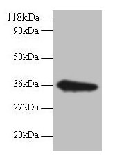
Figure 1. Western blot analysis of TSTA3/GFUS using anti-TSTA3/GFUS antibody (A09921-3). Electrophoresis was performed on a 5-20% SDS-PAGE gel at 70V (Stacking gel) / 90V (Resolving gel) for 2-3 hours. The sample well of each lane was loaded with 30 ug of sample under reducing conditions. Lane 1: human MCF-7 whole cell lysates, Lane 2: human Hela whole cell lysates, Lane 3: human HepG2 whole cell lysates, Lane 4: human RT4 whole cell lysates, Lane 5: human PC-3 whole cell lysates, Lane 6: rat testis tissue lysates, Lane 7: rat kidney tissue lysates, Lane 8: rat brain tissue lysates, Lane 9: mouse testis tissue lysates, Lane 10: mouse kidney tissue lysates. After electrophoresis, proteins were transferred to a nitrocellulose membrane at 150 mA for 50-90 minutes. Blocked the membrane with 5% non-fat milk/TBS for 1.5 hour at RT. The membrane was incubated with rabbit anti-TSTA3/GFUS antigen affinity purified polyclonal antibody (Catalog # A09921-3) at 0.25 microg/mL overnight at 4°C, then washed with TBS-0.1%Tween 3 times with 5 minutes each and probed with a goat anti-rabbit IgG-HRP secondary antibody at a dilution of 1:5000 for 1.5 hour at RT. The signal is developed using an Enhanced Chemiluminescent detection (ECL) kit (Catalog # EK1002) with Tanon 5200 system. A specific band was detected for TSTA3/GFUS at approximately 39 kDa. The expected band size for TSTA3/GFUS is at 39 kDa.
. TSTA3/GFUS was detected in an immunocytochemical section of MCF-7 cells. Enzyme antigen retrieval was performed using IHC enzyme antigen retrieval reagent (AR0022) for 15 mins. The cells were blocked with 10% goat serum. And then incubated with 5 microg/mL rabbit anti-TSTA3/GFUS Antibody (A09921-3) overnight at 4°C. DyLight®488 Conjugated Goat Anti-Rabbit IgG (BA1127) was used as secondary antibody at 1:100 dilution and incubated for 30 minutes at 37°C. The section was counterstained with DAPI. Visualize using a fluorescence microscope and filter sets appropriate for the label used.")
. Overlay histogram showing U87 cells stained with A09921-3 (Blue line). To facilitate intracellular staining, cells were fixed with 4% paraformaldehyde and permeabilized with permeabilization buffer. The cells were blocked with 10% normal goat serum. And then incubated with rabbit anti-TSTA3/GFUS Antibody (A09921-3, 1 microg/1x106 cells) for 30 min at 20°C. DyLight®488 conjugated goat anti-rabbit IgG (BA1127, 5-10 microg/1x106 cells) was used as secondary antibody for 30 minutes at 20°C. Isotype control antibody (Green line) was rabbit IgG (1 microg/1x106) used under the same conditions. Unlabelled sample without incubation with primary antibody and secondary antibody (Red line) was used as a blank control.")
Figure 1. Western blot analysis of TSTA3/GFUS using anti-TSTA3/GFUS antibody (A09921-3). Electrophoresis was performed on a 5-20% SDS-PAGE gel at 70V (Stacking gel) / 90V (Resolving gel) for 2-3 hours. The sample well of each lane was loaded with 30 ug of sample under reducing conditions. Lane 1: human MCF-7 whole cell lysates, Lane 2: human Hela whole cell lysates, Lane 3: human HepG2 whole cell lysates, Lane 4: human RT4 whole cell lysates, Lane 5: human PC-3 whole cell lysates, Lane 6: rat testis tissue lysates, Lane 7: rat kidney tissue lysates, Lane 8: rat brain tissue lysates, Lane 9: mouse testis tissue lysates, Lane 10: mouse kidney tissue lysates. After electrophoresis, proteins were transferred to a nitrocellulose membrane at 150 mA for 50-90 minutes. Blocked the membrane with 5% non-fat milk/TBS for 1.5 hour at RT. The membrane was incubated with rabbit anti-TSTA3/GFUS antigen affinity purified polyclonal antibody (Catalog # A09921-3) at 0.25 microg/mL overnight at 4°C, then washed with TBS-0.1%Tween 3 times with 5 minutes each and probed with a goat anti-rabbit IgG-HRP secondary antibody at a dilution of 1:5000 for 1.5 hour at RT. The signal is developed using an Enhanced Chemiluminescent detection (ECL) kit (Catalog # EK1002) with Tanon 5200 system. A specific band was detected for TSTA3/GFUS at approximately 39 kDa. The expected band size for TSTA3/GFUS is at 39 kDa.
Anti-TSTA3/GFUS Antibody Picoband(r)
A09921-3-CARRIER-FREE
ApplicationsFlow Cytometry, ImmunoFluorescence, Western Blot, ELISA, ImmunoCytoChemistry
Product group Antibodies
ReactivityHuman, Mouse, Rat
TargetGFUS
Overview
- SupplierBoster Bio
- Product NameAnti-TSTA3/GFUS Antibody Picoband(r)
- Delivery Days Customer9
- ApplicationsFlow Cytometry, ImmunoFluorescence, Western Blot, ELISA, ImmunoCytoChemistry
- CertificationResearch Use Only
- ClonalityPolyclonal
- Concentration500 ug/ml
- Gene ID7264
- Target nameGFUS
- Target descriptionGDP-L-fucose synthase
- Target synonymsFX, P35B, SDR4E1, TSTA3, GDP-L-fucose synthase, 3-5 epimerase/4-reductase, GDP-4-keto-6-deoxy-D-mannose epimerase-reductase, GDP-4-keto-6-deoxy-D-mannose-3,5-epimerase-4-reductase, red cell NADP(H)-binding protein, short chain dehydrogenase/reductase family 4E, member 1, testis tissue sperm-binding protein Li 45a, tissue specific transplantation antigen 3, tissue specific transplantation antigen P35B
- HostRabbit
- IsotypeIgG
- Protein IDQ13630
- Protein NameGDP-L-fucose synthase
- Scientific DescriptionBoster Bio Anti-TSTA3/GFUS Antibody Picoband® catalog # A09921-3. Tested in ELISA, Flow Cytometry, IF, ICC, WB applications. This antibody reacts with Human, Mouse, Rat. The brand Picoband indicates this is a premium antibody that guarantees superior quality, high affinity, and strong signals with minimal background in Western blot applications. Only our best-performing antibodies are designated as Picoband, ensuring unmatched performance.
- ReactivityHuman, Mouse, Rat
- Storage Instruction-20°C,2°C to 8°C
- UNSPSC12352203
Related products
Product group Antibodies
TSTA3 AntibodyCSB-PA00935A0RB
ApplicationsImmunoFluorescence, Western Blot, ELISA, ImmunoHistoChemistry
ReactivityHuman
TargetGFUS
- SizePrice
Product group Antibodies
Anti-TSTA3 AntibodyA97129
ApplicationsWestern Blot, ELISA
ReactivityHuman, Mouse, Rat
- SizePrice
Product group Antibodies

TSTA3 / GDP-L-Fucose Synthase AntibodyLS-C830985
ApplicationsELISA, ImmunoHistoChemistry
ReactivityHuman, Mouse
TargetGFUS
- SizePrice

Product group Antibodies
Anti-TSTA3 AntibodyHPA023361
ApplicationsWestern Blot, ImmunoCytoChemistry, ImmunoHistoChemistry
ReactivityHuman
TargetGFUS
- SizePrice
Product group Antibodies
TSTA3 Polyclonal AntibodyCAC13736
ApplicationsImmunoFluorescence, Western Blot, ELISA, ImmunoHistoChemistry
TargetGFUS
- SizePrice
Product group Antibodies
TSTA3 antibody [C1C3]GTX101663
ApplicationsWestern Blot
ReactivityHamster, Human
TargetGFUS
- SizePrice
Product group Antibodies
Anti-Mouse Tsta3 (C-term) Antibody102-23574
ApplicationsWestern Blot
TargetGFUS
- SizePrice