

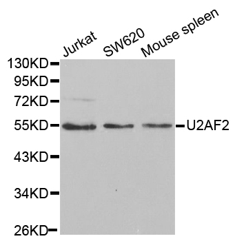

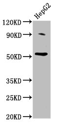
Anti-U2AF2 Antibody
A30527
ApplicationsImmunoFluorescence, Western Blot, ImmunoHistoChemistry
Product group Antibodies
ReactivityHuman, Mouse, Rat
Overview
- SupplierAntibodies.com
- Product NameAnti-U2AF2 Antibody
- Delivery Days Customer7
- ApplicationsImmunoFluorescence, Western Blot, ImmunoHistoChemistry
- CertificationResearch Use Only
- ClonalityPolyclonal
- ConjugateUnconjugated
- Estimated Purity>95%
- HostRabbit
- Scientific DescriptionRabbit polyclonal antibody to U2AF2
- ReactivityHuman, Mouse, Rat
- UNSPSC12352203
Related products

Product group Antibodies
Anti-U2AF65/U2AF2 Antibody Picoband(r)A03639-2-CARRIER-FREE
ApplicationsFlow Cytometry, ImmunoFluorescence, Western Blot, ELISA, ImmunoCytoChemistry, ImmunoHistoChemistry
ReactivityHuman, Mouse, Rat
TargetU2AF2
- SizePrice
Product group Antibodies
Anti-U2AF2 Antibody144-01936
ApplicationsImmunoFluorescence, ImmunoPrecipitation, Western Blot
ReactivityHuman, Mouse
TargetU2AF2
- SizePrice
Product group Antibodies
U2AF2 Recombinant AntibodyBSM-62036R
ApplicationsImmunoFluorescence, Western Blot, ImmunoCytoChemistry, ImmunoHistoChemistry, ImmunoHistoChemistry Frozen, ImmunoHistoChemistry Paraffin
ReactivityHuman, Mouse, Rat
TargetU2AF2
- SizePrice
Product group Antibodies
U2AF2 Polyclonal AntibodyCAC15375
ApplicationsWestern Blot, ELISA
TargetU2AF2
- SizePrice
Product group Antibodies
U2AF2 AntibodyCSB-PA025408LA01HU
ApplicationsWestern Blot, ELISA
ReactivityHuman
TargetU2AF2
- SizePrice
Product group Antibodies

U2AF2 / U2AF65 AntibodyLS-C331774
ApplicationsImmunoFluorescence, ImmunoPrecipitation, Western Blot, ImmunoHistoChemistry
ReactivityHuman, Mouse
TargetU2AF2
- SizePrice

Product group Antibodies
Anti-U2AF2 AntibodyHPA041943
ApplicationsWestern Blot, ImmunoCytoChemistry
ReactivityHuman
TargetU2AF2
- SizePrice
![U2AF65 antibody [C1C3] detects U2AF65 protein at cytoplasm and nucleus by immunohistochemical analysis. Sample: Paraffin-embedded rat colon. U2AF65 stained by U2AF65 antibody [C1C3] (GTX115622) diluted at 1:500. Antigen Retrieval: Citrate buffer, pH 6.0, 15 min](https://www.genetex.com/upload/website/prouct_img/normal/GTX115622/GTX115622_44518_20230414_IHC-P_R_23041023_586.webp)
Product group Antibodies
U2AF65 antibody [C1C3]GTX115622
ApplicationsImmunoFluorescence, Western Blot, ImmunoCytoChemistry, ImmunoHistoChemistry, ImmunoHistoChemistry Paraffin
ReactivityHuman, Mouse, Rat
TargetU2AF2
- SizePrice
Product group Antibodies
Anti-U2AF2/U2AF65 AntibodyCAB1936
ApplicationsImmunoFluorescence, ImmunoPrecipitation, Western Blot, ELISA, ImmunoCytoChemistry
ReactivityHuman
TargetU2AF2
- SizePrice