


Figure 1. Western blot analysis of Vitronectin using anti-Vitronectin antibody (PB9453). Electrophoresis was performed on a 5-20% SDS-PAGE gel at 70V (Stacking gel) / 90V (Resolving gel) for 2-3 hours. Lane 1: Rat Brain Tissue Lysate at 50ug, Lane 2: HEPA Whole Cell Lysate at 40ug. After electrophoresis, proteins were transferred to a nitrocellulose membrane at 150 mA for 50-90 minutes. Blocked the membrane with 5% non-fat milk/TBS for 1.5 hour at RT. The membrane was incubated with rabbit anti-Vitronectin antigen affinity purified polyclonal antibody (Catalog # PB9453) at 0.5 microg/mL overnight at 4°C, then washed with TBS-0.1%Tween 3 times with 5 minutes each and probed with a goat anti-rabbit IgG-HRP secondary antibody at a dilution of 1:5000 for 1.5 hour at RT. The signal is developed using an Enhanced Chemiluminescent detection (ECL) kit (Catalog # EK1002) with Tanon 5200 system. A specific band was detected for Vitronectin at approximately 373 kDa. The expected band size for Vitronectin is at 373 kDa.
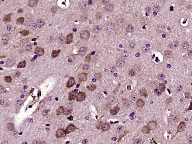
. Vitronectin was detected in a paraffin-embedded section of human glioma tissue. Heat mediated antigen retrieval was performed in EDTA buffer (pH 8.0, epitope retrieval solution). The tissue section was blocked with 10% goat serum. The tissue section was then incubated with 1 microg/ml rabbit anti-Vitronectin Antibody (PB9453) overnight at 4°C. Biotinylated goat anti-rabbit IgG was used as secondary antibody and incubated for 30 minutes at 37°C. The tissue section was developed using Strepavidin-Biotin-Complex (SABC) (Catalog # SA1022) with DAB as the chromogen.")
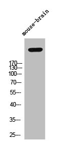
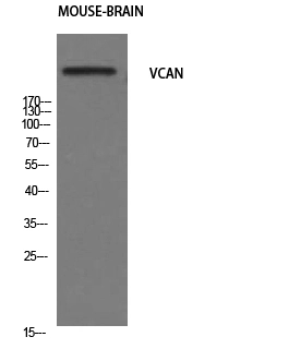
. Electrophoresis was performed on a 5-20% SDS-PAGE gel at 70V (Stacking gel) / 90V (Resolving gel) for 2-3 hours. The sample well of each lane was loaded with 30 ug of sample under reducing conditions. After electrophoresis, proteins were transferred to a nitrocellulose membrane at 150 mA for 50-90 minutes. Blocked the membrane with 5% milk with TBST for 1.5 hour at RT. The membrane was incubated with rabbit anti-versican/VCAN Antibody (PB9453) at 1: 500 overnight at 4°C , then washed with TBS-0.1%Tween 3 times with 5 minutes each and probed with an anti-rabbit 1 hour at room temperature. The signal is developed using an Enhanced Chemiluminescent detection (ECL) kit (Catalog # EK1002) with Tanon 5200 system. A specific band was detected for versican/VCAN at approximately 245 kDa. The expected band size for versican/VCAN is at 373 kDa.")
Figure 1. Western blot analysis of Vitronectin using anti-Vitronectin antibody (PB9453). Electrophoresis was performed on a 5-20% SDS-PAGE gel at 70V (Stacking gel) / 90V (Resolving gel) for 2-3 hours. Lane 1: Rat Brain Tissue Lysate at 50ug, Lane 2: HEPA Whole Cell Lysate at 40ug. After electrophoresis, proteins were transferred to a nitrocellulose membrane at 150 mA for 50-90 minutes. Blocked the membrane with 5% non-fat milk/TBS for 1.5 hour at RT. The membrane was incubated with rabbit anti-Vitronectin antigen affinity purified polyclonal antibody (Catalog # PB9453) at 0.5 microg/mL overnight at 4°C, then washed with TBS-0.1%Tween 3 times with 5 minutes each and probed with a goat anti-rabbit IgG-HRP secondary antibody at a dilution of 1:5000 for 1.5 hour at RT. The signal is developed using an Enhanced Chemiluminescent detection (ECL) kit (Catalog # EK1002) with Tanon 5200 system. A specific band was detected for Vitronectin at approximately 373 kDa. The expected band size for Vitronectin is at 373 kDa.
Anti-Versican/VCAN Antibody Picoband(r)
PB9453-CARRIER-FREE
ApplicationsWestern Blot, ImmunoHistoChemistry
Product group Antibodies
ReactivityHuman, Mouse, Rat
TargetVCAN
Overview
- SupplierBoster Bio
- Product NameAnti-Versican/VCAN Antibody Picoband(r)
- Delivery Days Customer9
- Application Supplier NoteWB: The detection limit for Versican is approximately 0.1ng/lane under reducing conditions. Tested Species: In-house tested species with positive results. Predicted Species: Species predicted to be fit for the product based on sequence similarities. By Heat: Boiling the paraffin sections in 10mM citrate buffer, pH6.0, for 20mins is required for the staining of formalin/paraffin sections. Other applications have not been tested. Optimal dilutions should be determined by end users.
- ApplicationsWestern Blot, ImmunoHistoChemistry
- CertificationResearch Use Only
- ClonalityPolyclonal
- Concentration500 ug/ml
- Gene ID1462
- Target nameVCAN
- Target descriptionversican
- Target synonymsCSPG2, ERVR, GHAP, PG-M, WGN, WGN1, versican core protein, chondroitin sulfate proteoglycan core protein 2, glial hyaluronate-binding protein, large fibroblast proteoglycan, versican proteoglycan
- HostRabbit
- IsotypeIgG
- Protein IDP13611
- Protein NameVersican core protein
- Scientific DescriptionBoster Bio Anti-Versican/VCAN Antibody Picoband® catalog # PB9453. Tested in IHC, WB applications. This antibody reacts with Human, Mouse, Rat. The brand Picoband indicates this is a premium antibody that guarantees superior quality, high affinity, and strong signals with minimal background in Western blot applications. Only our best-performing antibodies are designated as Picoband, ensuring unmatched performance.
- ReactivityHuman, Mouse, Rat
- Storage Instruction-20°C,2°C to 8°C
- UNSPSC12352203
Related products
Product group Antibodies
VCAN AntibodyCSB-PA007374
ApplicationsWestern Blot, ELISA, ImmunoHistoChemistry
ReactivityHuman
TargetVCAN
- SizePrice

Product group Antibodies
Anti-VCAN AntibodyA101248
ApplicationsELISA, ImmunoHistoChemistry
ReactivityHuman
- SizePrice

Product group Antibodies
Anti-VCAN AntibodyHPA004726
ApplicationsImmunoHistoChemistry
ReactivityHuman
TargetVCAN
- SizePrice
Product group Antibodies

ApplicationsWestern Blot, ELISA, ImmunoHistoChemistry
ReactivityHuman
TargetVCAN
- SizePrice
Product group Antibodies
ApplicationsWestern Blot, ImmunoHistoChemistry
TargetVCAN
- SizePrice
Product group Antibodies
Versican antibodyGTX34292
ApplicationsWestern Blot
ReactivityHuman, Mouse
TargetVCAN
- SizePrice
Product group Antibodies
Anti-VCAN Antibody130-10766
ApplicationsELISA
ReactivityHuman
TargetVCAN
- SizePrice
Product group Antibodies
Versican Polyclonal AntibodyBS-2533R
ApplicationsImmunoFluorescence, ELISA, ImmunoCytoChemistry, ImmunoHistoChemistry, ImmunoHistoChemistry Frozen, ImmunoHistoChemistry Paraffin
ReactivityBovine, Canine, Equine, Human, Mouse, Porcine, Rat
TargetVCAN
- SizePrice