


Figure 1. Western blot analysis of XBP using anti-XBP antibody (PB9463). Electrophoresis was performed on a 5-20% SDS-PAGE gel at 70V (Stacking gel) / 90V (Resolving gel) for 2-3 hours. Lane 1: MCF-7 Whole Cell Lysate at 40ug, Lane 2: MM231 Whole Cell Lysate at 40ug, Lane 3: MM453 Whole Cell Lysate at 40ug, Lane 4: SKOV Whole Cell Lysate at 40ug, Lane 5: HELA Whole Cell Lysate at 40ug. After electrophoresis, proteins were transferred to a nitrocellulose membrane at 150 mA for 50-90 minutes. Blocked the membrane with 5% non-fat milk/TBS for 1.5 hour at RT. The membrane was incubated with rabbit anti-XBP antigen affinity purified polyclonal antibody (Catalog # PB9463) at 0.5 microg/mL overnight at 4°C, then washed with TBS-0.1%Tween 3 times with 5 minutes each and probed with a goat anti-rabbit IgG-HRP secondary antibody at a dilution of 1:5000 for 1.5 hour at RT. The signal is developed using an Enhanced Chemiluminescent detection (ECL) kit (Catalog # EK1002) with Tanon 5200 system. A specific band was detected for XBP at approximately 29 kDa. The expected band size for XBP is at 29 kDa.
. XBP was detected in a paraffin-embedded section of mouse lung tissue. Heat mediated antigen retrieval was performed in EDTA buffer (pH 8.0, epitope retrieval solution). The tissue section was blocked with 10% goat serum. The tissue section was then incubated with 1 microg/ml rabbit anti-XBP Antibody (PB9463) overnight at 4°C. Biotinylated goat anti-rabbit IgG was used as secondary antibody and incubated for 30 minutes at 37°C. The tissue section was developed using Strepavidin-Biotin-Complex (SABC) (Catalog # SA1022) with DAB as the chromogen.")
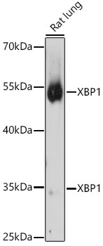
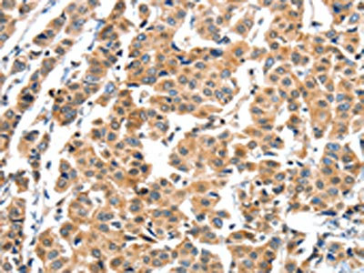
. XBP was detected in a paraffin-embedded section of rat lung tissue. Heat mediated antigen retrieval was performed in EDTA buffer (pH 8.0, epitope retrieval solution). The tissue section was blocked with 10% goat serum. The tissue section was then incubated with 1 microg/ml rabbit anti-XBP Antibody (PB9463) overnight at 4°C. Biotinylated goat anti-rabbit IgG was used as secondary antibody and incubated for 30 minutes at 37°C. The tissue section was developed using Strepavidin-Biotin-Complex (SABC) (Catalog # SA1022) with DAB as the chromogen.")
. XBP was detected in a paraffin-embedded section of human tonsil tissue. Heat mediated antigen retrieval was performed in EDTA buffer (pH 8.0, epitope retrieval solution). The tissue section was blocked with 10% goat serum. The tissue section was then incubated with 1 microg/ml rabbit anti-XBP Antibody (PB9463) overnight at 4°C. Biotinylated goat anti-rabbit IgG was used as secondary antibody and incubated for 30 minutes at 37°C. The tissue section was developed using Strepavidin-Biotin-Complex (SABC) (Catalog # SA1022) with DAB as the chromogen.")
. Overlay histogram showing HepG2 cells stained with PB9463 (Blue line). To facilitate intracellular staining, cells were fixed with 4% paraformaldehyde and permeabilized with permeabilization buffer. The cells were blocked with 10% normal goat serum. And then incubated with rabbit anti-XBP1 Antibody (PB9463, 1microg/1x106 cells) for 30 min at 20°C. DyLight®488 conjugated goat anti-rabbit IgG (BA1127, 5-10microg/1x106 cells) was used as secondary antibody for 30 minutes at 20°C. Isotype control antibody (Green line) was rabbit IgG (1microg/1x106) used under the same conditions. Unlabelled sample without incubation with primary antibody and secondary antibody (Red line) was used as a blank control.")
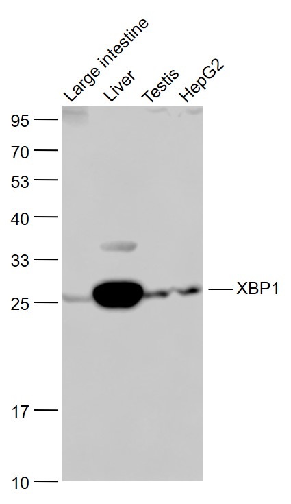
Figure 1. Western blot analysis of XBP using anti-XBP antibody (PB9463). Electrophoresis was performed on a 5-20% SDS-PAGE gel at 70V (Stacking gel) / 90V (Resolving gel) for 2-3 hours. Lane 1: MCF-7 Whole Cell Lysate at 40ug, Lane 2: MM231 Whole Cell Lysate at 40ug, Lane 3: MM453 Whole Cell Lysate at 40ug, Lane 4: SKOV Whole Cell Lysate at 40ug, Lane 5: HELA Whole Cell Lysate at 40ug. After electrophoresis, proteins were transferred to a nitrocellulose membrane at 150 mA for 50-90 minutes. Blocked the membrane with 5% non-fat milk/TBS for 1.5 hour at RT. The membrane was incubated with rabbit anti-XBP antigen affinity purified polyclonal antibody (Catalog # PB9463) at 0.5 microg/mL overnight at 4°C, then washed with TBS-0.1%Tween 3 times with 5 minutes each and probed with a goat anti-rabbit IgG-HRP secondary antibody at a dilution of 1:5000 for 1.5 hour at RT. The signal is developed using an Enhanced Chemiluminescent detection (ECL) kit (Catalog # EK1002) with Tanon 5200 system. A specific band was detected for XBP at approximately 29 kDa. The expected band size for XBP is at 29 kDa.
Anti-XBP1 Antibody Picoband(r)
PB9463
ApplicationsFlow Cytometry, ImmunoFluorescence, Western Blot, ImmunoCytoChemistry, ImmunoHistoChemistry
Product group Antibodies
ReactivityHamster, Human, Mouse, Rat
TargetXBP1
Overview
- SupplierBoster Bio
- Product NameAnti-XBP1 Antibody Picoband(r)
- Delivery Days Customer9
- Application Supplier NoteWB: The detection limit for XBP1 is approximately 0.1ng/lane under reducing conditions. Tested Species: In-house tested species with positive results. By Heat: Boiling the paraffin sections in 10mM citrate buffer, pH6.0, for 20mins is required for the staining of formalin/paraffin sections. Other applications have not been tested. Optimal dilutions should be determined by end users.
- ApplicationsFlow Cytometry, ImmunoFluorescence, Western Blot, ImmunoCytoChemistry, ImmunoHistoChemistry
- CertificationResearch Use Only
- ClonalityPolyclonal
- Concentration500 ug/ml
- Gene ID7494
- Target nameXBP1
- Target descriptionX-box binding protein 1
- Target synonymsTREB-5, TREB5, XBP-1, XBP2, X-box-binding protein 1, tax-responsive element-binding protein 5
- HostRabbit
- IsotypeIgG
- Protein IDP17861
- Protein NameX-box-binding protein 1
- Scientific DescriptionBoster Bio Anti-XBP1 Antibody Picoband® catalog # PB9463. Tested in Flow Cytometry, IF, IHC, ICC, WB applications. This antibody reacts with Human, Mouse, Rat. The brand Picoband indicates this is a premium antibody that guarantees superior quality, high affinity, and strong signals with minimal background in Western blot applications. Only our best-performing antibodies are designated as Picoband, ensuring unmatched performance.
- ReactivityHamster, Human, Mouse, Rat
- Storage Instruction-20°C,2°C to 8°C
- UNSPSC12352203
References
- Li C, Ma Y, Chai X, et al. Ketogenic diet attenuates cognitive dysfunctions induced by hypoglycemia via inhibiting endoplasmic reticulum stress-dependent pathways. Food Funct. 2024,15(3):1294-1309. doi: 10.1039/d3fo04007kRead this paper
- Wang D, Si D, Li G, et al. Dysregulated autophagic activity induced in response to chronic intermittent hypoxia contributes to the pathogenesis of NAFLD. Front Physiol. 2022,13:941706. doi: 10.3389/fphys.2022.941706Read this paper
- Hu X, Zhang Q, Guo M, et al. Deletion of RNF186 expression suppresses diet-induced hepatic steatosis by regulating insulin activity. iScience. 2022,25(2):103859. doi: 10.1016/j.isci.2022.103859Read this paper
- Wei W, Gao Y, Wang C, et al. Excessive fluoride induces endoplasmic reticulum stress and interferes enamel proteinases secretion. Environ Toxicol. 2013,28(6):332-41. doi: 10.1002/tox.20724Read this paper
Datasheet
MSDS
Related products
Product group Antibodies
Anti-XBP1 Antibody144-01731
ApplicationsImmunoFluorescence, Western Blot
ReactivityHuman, Mouse, Rat
TargetXBP1
- SizePrice
Product group Antibodies
Anti-XBP1 AntibodyA13620
ApplicationsImmunoFluorescence, Western Blot, ImmunoCytoChemistry
ReactivityHuman, Mouse, Rat
- SizePrice
Product group Antibodies
XBP1 AntibodyCSB-PA027191
ApplicationsELISA, ImmunoHistoChemistry
ReactivityHuman
TargetXBP1
- SizePrice
![XBP1 antibody [N3C3] detects XBP1 protein at nucleus in mouse brain by immunohistochemical analysis. Sample: Paraffin-embedded mouse brain. XBP1 antibody [N3C3] (GTX102229) diluted at 1:500.
Antigen Retrieval: Citrate buffer, pH 6.0, 15 min](https://www.genetex.com/upload/website/prouct_img/normal/GTX102229/GTX102229_40079_20151130_IHC-P_M_w_23060100_471.webp)
Product group Antibodies
XBP1 antibody [N3C3]GTX102229
ApplicationsImmunoFluorescence, Western Blot, ImmunoCytoChemistry, ImmunoHistoChemistry, ImmunoHistoChemistry Paraffin
ReactivityHuman, Mouse, Rat
TargetXBP1
- SizePrice
Product group Antibodies

Goat anti-XBP1 / TREB5EB08557
ApplicationsImmunoFluorescence, Western Blot, ELISA, ImmunoHistoChemistry
ReactivityCanine, Human, Mouse, Rat
TargetXBP1
- SizePrice
Product group Antibodies

XBP1 AntibodyLS-C401118
ApplicationsELISA, ImmunoHistoChemistry
ReactivityHuman
TargetXBP1
- SizePrice
Product group Antibodies
Anti-XBP1 Antibody Picoband(r)PB9463-CARRIER-FREE
ApplicationsFlow Cytometry, ImmunoFluorescence, Western Blot, ImmunoCytoChemistry, ImmunoHistoChemistry
ReactivityHamster, Human, Mouse, Rat
TargetXBP1
- SizePrice
Product group Antibodies
References
XBP1 Polyclonal AntibodyBS-1668R
ApplicationsFlow Cytometry, ImmunoFluorescence, Western Blot, ELISA, ImmunoCytoChemistry, ImmunoHistoChemistry, ImmunoHistoChemistry Frozen, ImmunoHistoChemistry Paraffin
ReactivityBovine, Chicken, Human, Mouse, Rat
TargetXBP1
- SizePrice