


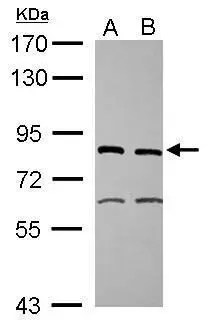
Figure 1. Western blot analysis of XPD/ERCC2 using anti-XPD/ERCC2 antibody (A00694-2). Electrophoresis was performed on a 5-20% SDS-PAGE gel at 70V (Stacking gel) / 90V (Resolving gel) for 2-3 hours. The sample well of each lane was loaded with 30 ug of sample under reducing conditions. Lane 1: human K562 whole cell lysates, Lane 2: human SiHa whole cell lysates, Lane 3: human HepG2 whole cell lysates. After electrophoresis, proteins were transferred to a nitrocellulose membrane at 150 mA for 50-90 minutes. Blocked the membrane with 5% non-fat milk/TBS for 1.5 hour at RT. The membrane was incubated with rabbit anti-XPD/ERCC2 antigen affinity purified polyclonal antibody (Catalog # A00694-2) at 0.5 microg/mL overnight at 4°C, then washed with TBS-0.1%Tween 3 times with 5 minutes each and probed with a goat anti-rabbit IgG-HRP secondary antibody at a dilution of 1:5000 for 1.5 hour at RT. The signal is developed using an Enhanced Chemiluminescent detection (ECL) kit (Catalog # EK1002) with Tanon 5200 system. A specific band was detected for XPD/ERCC2 at approximately 87 kDa. The expected band size for XPD/ERCC2 is at 80 kDa.
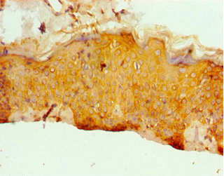
. XPD/ERCC2 was detected in a paraffin-embedded section of human bladder epithelial carcinom tissue. Heat mediated antigen retrieval was performed in EDTA buffer (pH 8.0, epitope retrieval solution). The tissue section was blocked with 10% goat serum. The tissue section was then incubated with 2 microg/ml rabbit anti-XPD/ERCC2 Antibody (A00694-2) overnight at 4°C. Biotinylated goat anti-rabbit IgG was used as secondary antibody and incubated for 30 minutes at 37°C. The tissue section was developed using Strepavidin-Biotin-Complex (SABC) (Catalog # SA1022) with DAB as the chromogen.")
. XPD/ERCC2 was detected in a paraffin-embedded section of mouse testis tissue. Heat mediated antigen retrieval was performed in EDTA buffer (pH 8.0, epitope retrieval solution). The tissue section was blocked with 10% goat serum. The tissue section was then incubated with 2 microg/ml rabbit anti-XPD/ERCC2 Antibody (A00694-2) overnight at 4°C. Biotinylated goat anti-rabbit IgG was used as secondary antibody and incubated for 30 minutes at 37°C. The tissue section was developed using Strepavidin-Biotin-Complex (SABC) (Catalog # SA1022) with DAB as the chromogen.")
. XPD/ERCC2 was detected in a paraffin-embedded section of rat testis tissue. Heat mediated antigen retrieval was performed in EDTA buffer (pH 8.0, epitope retrieval solution). The tissue section was blocked with 10% goat serum. The tissue section was then incubated with 2 microg/ml rabbit anti-XPD/ERCC2 Antibody (A00694-2) overnight at 4°C. Biotinylated goat anti-rabbit IgG was used as secondary antibody and incubated for 30 minutes at 37°C. The tissue section was developed using Strepavidin-Biotin-Complex (SABC) (Catalog # SA1022) with DAB as the chromogen.")
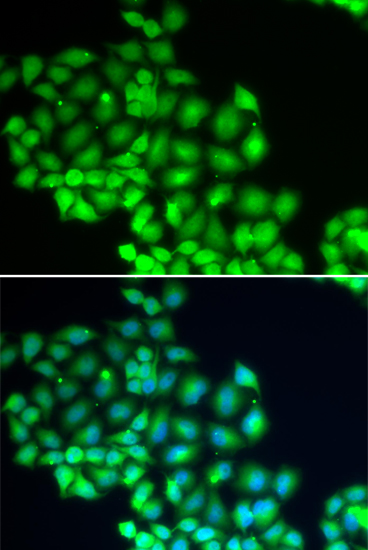
. Overlay histogram showing THP-1 cells stained with A00694-2 (Blue line). To facilitate intracellular staining, cells were fixed with 4% paraformaldehyde and permeabilized with permeabilization buffer. The cells were blocked with 10% normal goat serum. And then incubated with rabbit anti-XPD/ERCC2 Antibody (A00694-2, 1 microg/1x106 cells) for 30 min at 20°C. DyLight®488 conjugated goat anti-rabbit IgG (BA1127, 5-10 microg/1x106 cells) was used as secondary antibody for 30 minutes at 20°C. Isotype control antibody (Green line) was rabbit IgG (1 microg/1x106) used under the same conditions. Unlabelled sample without incubation with primary antibody and secondary antibody (Red line) was used as a blank control.")
Figure 1. Western blot analysis of XPD/ERCC2 using anti-XPD/ERCC2 antibody (A00694-2). Electrophoresis was performed on a 5-20% SDS-PAGE gel at 70V (Stacking gel) / 90V (Resolving gel) for 2-3 hours. The sample well of each lane was loaded with 30 ug of sample under reducing conditions. Lane 1: human K562 whole cell lysates, Lane 2: human SiHa whole cell lysates, Lane 3: human HepG2 whole cell lysates. After electrophoresis, proteins were transferred to a nitrocellulose membrane at 150 mA for 50-90 minutes. Blocked the membrane with 5% non-fat milk/TBS for 1.5 hour at RT. The membrane was incubated with rabbit anti-XPD/ERCC2 antigen affinity purified polyclonal antibody (Catalog # A00694-2) at 0.5 microg/mL overnight at 4°C, then washed with TBS-0.1%Tween 3 times with 5 minutes each and probed with a goat anti-rabbit IgG-HRP secondary antibody at a dilution of 1:5000 for 1.5 hour at RT. The signal is developed using an Enhanced Chemiluminescent detection (ECL) kit (Catalog # EK1002) with Tanon 5200 system. A specific band was detected for XPD/ERCC2 at approximately 87 kDa. The expected band size for XPD/ERCC2 is at 80 kDa.
Anti-XPD/ERCC2 Antibody Picoband(r)
A00694-2-CARRIER-FREE
ApplicationsFlow Cytometry, Western Blot, ELISA, ImmunoHistoChemistry
Product group Antibodies
ReactivityHuman, Mouse, Rat
TargetERCC2
Overview
- SupplierBoster Bio
- Product NameAnti-XPD/ERCC2 Antibody Picoband(r)
- Delivery Days Customer9
- ApplicationsFlow Cytometry, Western Blot, ELISA, ImmunoHistoChemistry
- CertificationResearch Use Only
- ClonalityPolyclonal
- Concentration500 ug/ml
- Gene ID2068
- Target nameERCC2
- Target descriptionERCC excision repair 2, TFIIH core complex helicase subunit
- Target synonymsCOFS2, CXPD, EM9, TFIIH, TTD, TTD1, XPD, general transcription and DNA repair factor IIH helicase subunit XPD, BTF2 p80, DNA 5'-3' helicase XPD, DNA excision repair protein ERCC-2, DNA repair protein complementing XP-D cells, TFIIH 80 kDa subunit, TFIIH basal transcription factor complex 80 kDa subunit, TFIIH basal transcription factor complex helicase XPD subunit, TFIIH p80, TFIIH subunit XPD, basic transcription factor 2 80 kDa subunit, excision repair cross-complementation group 2, excision repair cross-complementing rodent repair deficiency, complementation group 2, xeroderma pigmentosum complementary group D, xeroderma pigmentosum group D-complementing protein
- HostRabbit
- IsotypeIgG
- Protein IDP18074
- Protein NameGeneral transcription and DNA repair factor IIH helicase subunit XPD
- Scientific DescriptionBoster Bio Anti-XPD/ERCC2 Antibody Picoband® catalog # A00694-2. Tested in ELISA, Flow Cytometry, IHC, WB applications. This antibody reacts with Human, Mouse, Rat. The brand Picoband indicates this is a premium antibody that guarantees superior quality, high affinity, and strong signals with minimal background in Western blot applications. Only our best-performing antibodies are designated as Picoband, ensuring unmatched performance.
- ReactivityHuman, Mouse, Rat
- Storage Instruction-20°C,2°C to 8°C
- UNSPSC12352203
Related products
Product group Antibodies
ERCC2 AntibodyCSB-PA007770ESR1HU
ApplicationsELISA, ImmunoHistoChemistry
ReactivityHuman
TargetERCC2
- SizePrice
Product group Antibodies

ERCC2 / XPD AntibodyLS-C749564
ApplicationsWestern Blot
ReactivityHuman, Mouse, Rat
TargetERCC2
- SizePrice
Product group Antibodies
Anti-ERCC2 AntibodyA39743
ApplicationsImmunoFluorescence, Western Blot
ReactivityHuman, Mouse, Rat
- SizePrice

Product group Antibodies
Anti-ERCC2 AntibodyHPA038057
ApplicationsWestern Blot, ImmunoHistoChemistry
ReactivityHuman
TargetERCC2
- SizePrice
Product group Antibodies
ERCC2 Polyclonal AntibodyCAC13030
ApplicationsWestern Blot, ELISA, ImmunoHistoChemistry
TargetERCC2
- SizePrice
Product group Antibodies
XPD antibody [N2C2], InternalGTX105357
ApplicationsImmunoFluorescence, Western Blot, ImmunoCytoChemistry, ImmunoHistoChemistry, ImmunoHistoChemistry Paraffin
ReactivityHuman, Mouse
TargetERCC2
- SizePrice
Product group Antibodies
XPD Recombinant Antibody, AbBy Fluor-405 ConjugatedBSM-62366R-BF405
ApplicationsImmunoFluorescence, Western Blot
ReactivityHuman
TargetERCC2
- SizePrice
Product group Antibodies
Anti-ERCC2 Antibody144-60710
ApplicationsWestern Blot
ReactivityHuman, Mouse, Rat
TargetERCC2
- SizePrice