Bio-Connect

Antibodies

We offer one of the most comprehensive portfolios of antibodies. This includes monoclonal and polyclonal primary, secondary, conjugated, phospho-specific, functional, (isotype) controls, tagged and antibody pairs. In addition, we offer custom antibody services from several manufacturers.

The antibodies are generated in various hosts and react to antigens of different species like human, mouse, rat, rabbit or zebrafish. The antibodies are validated for multiple applications, including immunohistochemistry, western blot, immunoprecipitation, ELISA and flow cytometry, to ensure reliable performance for your research needs.

If you need a specific antibody and can’t find it in our webshop, please contact our technical support.

Discover what our customers say about us by reading their reviews.

1045 to 1056 of 90.124 products
Sort by
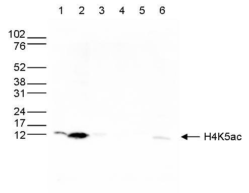
Western blot was performed on whole cell (25μg, lane 1) and histone extracts (15μg, lane 2) from HeLa cells, and on 1 μg of recombinant histone H2A, H2B, H3 and H4 (lane 3, 4, 5 and 6, respectively) using H4K5ac Polyclonal Antibody (bs-53099R). Antibody was diluted 1:500 in TBS-Tween containing 5% skim milk.
Product group Antibodies
Bioss Antibodies
ApplicationsDot Blot, ImmunoFluorescence, Western Blot, ELISA, ImmunoCytoChemistry, ImmunoHistoChemistry, ImmunoHistoChemistry Paraffin
ReactivityHuman, Mouse
TargetH4C9
  • SizePrice
HeLa cells were stained with the Bioss antibody against WDR5 (Cat. No. bs-53101R) and with DAPI. Cells were fixed with 4% formaldehyde for 10’ and blocked with PBS/TX-100 containing 5% normal goat serum and 1% BSA. The cells were immunofluorescently labeled with the WDR5 antibody (left) diluted 1:1,000 in blocking solution followed by an anti-rabbit antibody conjugated to Alexa488. The middle panel shows staining of the nuclei with DAPI. A merge of the two stainings is shown on the right.
Product group Antibodies
Bioss Antibodies
ApplicationsImmunoFluorescence, Western Blot, ChIP Chromatin ImmunoPrecipitation
ReactivityHuman
TargetWDR5
  • SizePrice
HeLa cells were stained with the Bioss antibody against H3K4me3 (Cat. No. bs-53103R) and with DAPI. Cells were fixed with 4% formaldehyde for 10’ and blocked with PBS/TX-100 containing 5% normal goat serum and 1% BSA. The cells were immunofluorescently labeled with the H3K4me3 antibody (left) diluted 1:100 in blocking solution followed by an anti-rabbit antibody conjugated to Alexa488. The middle panel shows staining of the nuclei with DAPI. A merge of the two stainings is shown on the right.
Product group Antibodies
Bioss Antibodies
ApplicationsDot Blot, ImmunoFluorescence, Western Blot, ChIP Chromatin ImmunoPrecipitation
ReactivityHuman, Mouse, Plant, Other Species
TargetH3C1
  • SizePrice
HeLa cells were stained with the Bioss antibody against H4K20me1 (Cat. bs-53104R) and with DAPI. Cells were fixed with 4% formaldehyde for 10’ and blocked with PBS/TX-100 containing 5% normal goat serum and 1% BSA. The cells were immunofluorescently labeled with the H4K20me1 antibody (left) diluted 1:500 in blocking solution followed by an anti-rabbit antibody conjugated to Alexa488. The middle panel shows staining of the nuclei with DAPI. A merge of the two stainings is shown on the right.
Product group Antibodies
Bioss Antibodies
ApplicationsDot Blot, ImmunoFluorescence, Western Blot, ELISA, ImmunoCytoChemistry
ReactivityHuman
TargetH4C9
  • SizePrice
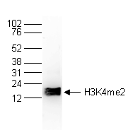
Histone extracts of HeLa cells (15 μg) were analyzed by Western blot using the Bioss antibody against H3K4me2 (Cat. No. bs-53105R) diluted 1:1,000 in TBS-Tween containing 5% skimmed milk. The position of the protein of interest is indicated on the right; the marker (in kDa) is shown on the left._x000D_
Product group Antibodies
Bioss Antibodies
ApplicationsDot Blot, Western Blot, ChIP Chromatin ImmunoPrecipitation, ELISA
ReactivityHuman
TargetH3C1
  • SizePrice
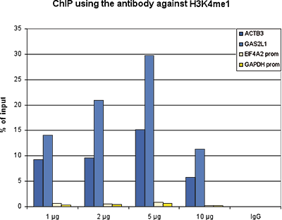
ChIP was performed with the Bioss antibody against H3K4me1 (Cat. No. bs-53106R) on sheared chromatin from 1 million HeLaS3 cells using a ChIP-seq kit. A titration of the antibody consisting of 1, 2, 5 and 10 μg per ChIP experiment was analyzed. IgG (2 μg/IP) was used as negative IP control. Quantitative PCR was performed with primers for a region surrounding the ACTB and GAS2L1 genes, used as positive controls, and for the promoters of the GAPDH and EIF4A2 genes, used as negative controls. The figure shows the recovery, expressed as a % of input (the relative amount of immunoprecipitated DNA compared to input DNA after qPCR analysis).
Product group Antibodies
Bioss Antibodies
ApplicationsDot Blot, ImmunoFluorescence, Western Blot, ChIP Chromatin ImmunoPrecipitation, ELISA
ReactivityHuman, Mouse
TargetH3C1
  • SizePrice
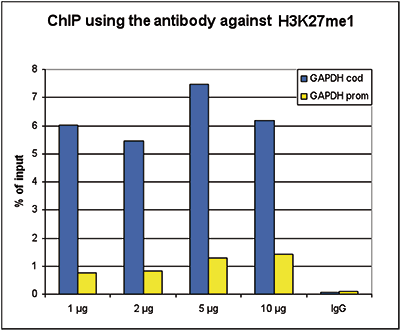
ChIP assays were performed using HeLa cells, the Bioss antibody against H3K27me1 (Cat. No. bs-53108R) and optimized PCR primer sets for qPCR. ChIP was performed using sheared chromatin from 100,000 cells. A titration of the antibody consisting of 1, 2, 5 and 10 μg per ChIP experiment was analyzed. IgG (2 μg/IP) was used as negative IP control. QPCR was performed with primers for the promoter and the coding region of the active gene GAPDH used as a negative and a positive control target, respectively. The figure shows the recovery, expressed as a % of input (the relative amount of immunoprecipitated DNA compared to input DNA after qPCR analysis).
Product group Antibodies
Bioss Antibodies
ApplicationsDot Blot, ImmunoFluorescence, Western Blot, ELISA
ReactivityHuman, Plant, Other Species
TargetH3C1
  • SizePrice
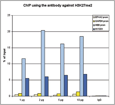
ChIP assays were performed using HeLa cells, the Bioss antibody against H3K27me2 (cat. No. bs-53109R) and optimized PCR primer sets for qPCR. ChIP was performed with an Auto Histone ChIP-seq kit on the SX-8G IP-Star Compact automated system, using sheared chromatin from 1 million cells. A titration of the antibody consisting of 1, 2, 5, and 10 μg per ChIP experiment was analyzed. IgG (2 μg/IP) was used as negative IP control. QPCR was performed with primers for the promoter of the active GAPDH and EIF4A2 genes, used as negative controls, and for the promoter of the inactive HBB and the coding region of the inactive MYOD1 genes, used as positive controls. The figure shows the recovery, expressed as a % of input (the relative amount of immunoprecipitated DNA compared to input DNA after qPCR analysis). These results are in accordance with the observation that H3K27me2 is preferably present at silent genes.
Product group Antibodies
Bioss Antibodies
ApplicationsDot Blot, ImmunoFluorescence, Western Blot, ELISA
ReactivityHuman
TargetH3C1
  • SizePrice
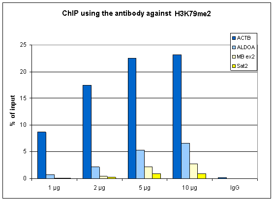
ChIP assays were performed using HeLa cells, the Bioss antibody against H3K79me2 (Cat. No. bs-53111R) and optimized PCR primer pairs for qPCR. ChIP was performed with an Auto Histone ChIP-seq kit, using sheared chromatin from 1 million cells. A titration consisting of 0.5, 1, 2 and 5 μg of antibody per ChIP experiment was analyzed. IgG (1 μg/IP) was used as a negative IP control. Quantitative PCR was performed with primers specific for the coding regions of the active EIF2S3 and CCT5 genes, used as positive controls, and for the inactive MYOD1) gene and the Sat2 satellite repeat, used as negative controls. The figure shows the recovery, expressed as a % of input (the relative amount of immunoprecipitated DNA compared to input DNA after qPCR analysis).
Product group Antibodies
Bioss Antibodies
ApplicationsDot Blot, Western Blot, ChIP Chromatin ImmunoPrecipitation, ELISA
ReactivityHuman
TargetH3C1
  • SizePrice
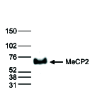
Nuclear extracts (40 μg) from HeLa cells were analysed by Western blot using the Bioss antibody against MeCP2 (Cat. No. bs-53112R) diluted 1:1,000 in TBS-Tween containing 5% skimmed milk. The position of the protein of interest is indicated on the right; the marker (in kDa) is shown on the left.
Product group Antibodies
Bioss Antibodies
ApplicationsDot Blot, Western Blot, ELISA
ReactivityHuman
TargetMECP2
  • SizePrice
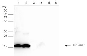
Western blot was performed on whole cell (25 µg, lane 1) and histone extracts (15 µg, lane 2) from HeLa cells, and on 1 µg of recombinant histone H2A, H2B, H3 and H4 (lane 3, 4, 5 and 6, respectively) using the Bioss antibody against H3K9me3 (Cat. No. bs-53114R). The antibody was diluted 1:1,000 in TBS-Tween containing 5% skimmed milk.
Product group Antibodies
Bioss Antibodies
ApplicationsDot Blot, ImmunoFluorescence, Western Blot, ELISA, ImmunoCytoChemistry
ReactivityHuman, Mouse, Plant, Zebra Fish, Other Species
TargetH3C1
  • SizePrice
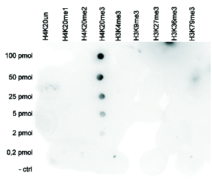
A Dot Blot analysis was performed to test the cross-reactivity of the Bioss antibody against H4K20me3 (Cat. No. bs-53115R) with peptides containing other histone modifications and the unmodified H4K20. One hundred to 0.2 pmol of the respective peptides were spotted on a membrane. The antibody was used at a dilution of 1:20,000. Figure shows a high specificity of the antibody for the modification of interest
Product group Antibodies
Bioss Antibodies
ApplicationsDot Blot, ImmunoFluorescence, Western Blot, ChIP Chromatin ImmunoPrecipitation, ELISA, ImmunoCytoChemistry, ImmunoHistoChemistry, ImmunoHistoChemistry Paraffin
ReactivityHuman, Mouse, Other Species
TargetH4C9
  • SizePrice