Bio-Connect

Antibodies

We offer one of the most comprehensive portfolios of antibodies. This includes monoclonal and polyclonal primary, secondary, conjugated, phospho-specific, functional, (isotype) controls, tagged and antibody pairs. In addition, we offer custom antibody services from several manufacturers.

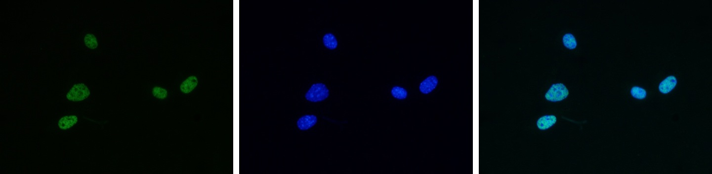
The antibodies are generated in various hosts and react to antigens of different species like human, mouse, rat, rabbit or zebrafish. The antibodies are validated for multiple applications, including immunohistochemistry, western blot, immunoprecipitation, ELISA and flow cytometry, to ensure reliable performance for your research needs.

If you need a specific antibody and can’t find it in our webshop, please contact our technical support.

Discover what our customers say about us by reading their reviews.

1033 to 1044 of 90.124 products
Sort by
To determine the titer of the antibody, an ELISA was performed using a serial dilution of the CDYL antibody (bs-53086R). The plates were coated with peptides. By plotting the absorbance against the antibody dilution, the titer of the antibody was estimated to be 1:8,700.
Product group Antibodies
Bioss Antibodies
ApplicationsELISA
ReactivityHuman, Mouse
TargetCDYL
  • SizePrice
To determine the titer of the antibody, an ELISA was performed using a serial dilution of the antibody directed against PIWIL1 (bs-53087R). The plates were coated with peptides. By plotting the absorbance against the antibody dilution, the titer of the antibody was estimated to be 1:23,800.
Product group Antibodies
Bioss Antibodies
ApplicationsELISA
ReactivityHuman
TargetPIWIL1
  • SizePrice
To determine the titer of the antibody, an ELISA was performed using a serial dilution of DGCR8 antibody (bs-53088R). The plates were coated with peptides. By plotting the absorbance against the antibody dilution, the titer of the antibody was estimated to be 1:8,100.
Product group Antibodies
Bioss Antibodies
ApplicationsELISA
ReactivityHuman, Mouse
TargetDGCR8
  • SizePrice
To determine the titer of the antibody, an ELISA was performed using a serial dilution of the H1.1 antibody (bs-53089R). The plates were coated with the peptide. By plotting the absorbance against the antibody dilution, the titer of the antibody was estimated to be 1:3,000.
Product group Antibodies
Bioss Antibodies
ApplicationsWestern Blot, ELISA
ReactivityHuman
TargetH1-1
  • SizePrice
To determine the titer of the antibody, an ELISA was performed using a serial dilution of the CBX4 antibody (bs-53090R). The plates were coated with peptides. By plotting the absorbance against the antibody dilution, the titer of the antibody was estimated to be 1:11,250.
Product group Antibodies
Bioss Antibodies
ApplicationsDot Blot, ELISA
ReactivityHuman
  • SizePrice
To determine the titer of the antibody, an ELISA was performed using a serial dilution MYH11 Polyclonal Antibody (bs-53091R). The plates were coated with the peptides. By plotting the absorbance against the antibody dilution, the titer of the antibody was estimated to be 1:1,900.
Product group Antibodies
Bioss Antibodies
ApplicationsWestern Blot, ChIP Chromatin ImmunoPrecipitation, ELISA
ReactivityHuman
TargetMYH11
  • SizePrice
ChIP assays were performed using HeLa cells, H3K9ac Polyclonal Antibody (Cat. No. bs-53092R) and optimized PCR primer pairs for qPCR. ChIP was performed with a ChIP-seq kit, using sheared chromatin from 1 million cells. A titration consisting of 1, 2, 5 and 10 μg of antibody per ChIP experiment was analyzed. IgG (2 μg/IP) was used as a negative IP control. Quantitative PCR was performed with primers specific for the promoter of the active genes GAPDH and EIF4A2, used as positive controls, and for exon 2 of the inactive myoglobin (MB) gene and the Sat2 satellite repeat, used as negative controls. The figure shows the recovery, expressed as a % of input (the relative amount of immunoprecipitated DNA compared to input DNA after qPCR analysis). These results are in accordance with the observation that acetylation of K9 at histone H3 is associated with the promoters of active genes.
Product group Antibodies
Bioss Antibodies
ApplicationsDot Blot, ImmunoFluorescence, Western Blot, ChIP Chromatin ImmunoPrecipitation, ELISA
ReactivityHuman, Mouse, Plant, Plasmodium, Other Species
TargetH3C1
  • SizePrice
ChIP assays were performed using HeLa cells, the Bioss antibody against H3K9/14ac (Cat. No. bs-53093R) and optimized primer pairs for qPCR. ChIP was performed with a HighCell# ChIP kit, using sheared chromatin from 1.5 million cells. A titration of the antibody consisting of 1, 2, 5 and 10 μg per ChIP experiment was analyzed. IgG (5 μg/IP) was used as negative IP control.QPCR was performed using primers specific for the promoter of the ACTB gene as a positive control target and for exon 2 of the MB gene as a negative control target. The figure shows the recovery (the relative amount of immunoprecipitated DNA compared to input DNA). These results confirm the observation that acetylation of H3K9/14 is present at active promoters.
Product group Antibodies
Bioss Antibodies
ApplicationsDot Blot, ImmunoFluorescence, Western Blot, ChIP Chromatin ImmunoPrecipitation, ELISA
ReactivityFungus, Human, Mouse, Plant, Other Species
TargetH3C1
  • SizePrice
Western blot was performed on HeLa nuclear extracts (lane 1) and purified human SKI complex (lane 2) (Figure A), or on nuclear extracts from HeLa cells (HeLa NE, 20 μg) (Figure B) using the Bioss antibody against SKI3 (Cat. No. bs-53094R) diluted 1:1,000.
Product group Antibodies
Bioss Antibodies
ApplicationsImmunoFluorescence, Western Blot, ImmunoCytoChemistry
ReactivityHuman
TargetSKI8
  • SizePrice
HeLa cells were stained with the Bioss antibody against H4K5,8,12ac (cat. No. bs-53096R) and with DAPI. Cells were fixed with 4% formaldehyde for 10’ and blocked with PBS/TX-100 containing 5% normal goat serum and 1% BSA. Figure A: cells were immunofluorescently labeled with the H4K5,8,12ac antibody (left) diluted 1:500 in blocking solution followed by Alexa488 conjugated secondary antibody. The middle panel shows staining of the nuclei with DAPI. A merge of the two stainings is shown on the right. Figure B, C, D, and E: staining of the cells with the H4K5,8,12ac antibody after incubation of the antibody with 10 ng/μl of the following blocking peptides: H4K5,8,12 unmodified (figure B), H4K5,8,12ac (figure C), H2A.ZK5,7,11ac (figure D) and H4K5,8,12,16ac (figure E).
Product group Antibodies
Bioss Antibodies
ApplicationsDot Blot, ImmunoFluorescence, ImmunoPrecipitation, Western Blot, ChIP Chromatin ImmunoPrecipitation, ELISA
ReactivityHuman, Mouse
TargetH4C9
  • SizePrice
Immunoprecipitation was performed on nuclear extracts from cells transfected with FLAG-tagged L3MBTL1 (L3MBTL1-F) with the Bioss antibody against L3MBTL1 (Cat. No. bs-53097R) and with an IgG negative control antibody. Western Blot was performed with an anti-FLAG antibody to demonstrate the presence of L3MBTL1-F in the input (lane 1), in the precipitated fraction (P) and in the supernatant (SN). These data show that L3MBTL1-F was efficiently precipitated by the L3MBTL1 antibody (lane 3 and 5), whereas it was not precipitated by the IgG negative control antibody (lane 2 and 4).
Product group Antibodies
Bioss Antibodies
ApplicationsImmunoFluorescence, ImmunoPrecipitation, Western Blot
ReactivityHuman
TargetL3MBTL1
  • SizePrice
Mouse NIH3T3 cells were stained with Bioss antibody against H4K5,8,12,16ac (cat. No. bs-53098R) and with DAPI. Cells were fixed with 4% formaldehyde for 10’ and blocked with PBS/TX-100 containing 5% normal goat serum and 1% BSA. The cells were immunofluorescently labeled with the H4K5,8,12,16ac antibody (left) diluted 1:500 in blocking solution followed by an anti-rabbit antibody conjugated to Alexa488. The middle panel shows staining of the nuclei with DAPI. A merge of the two stainings is shown on the right.
Product group Antibodies
Bioss Antibodies
ApplicationsDot Blot, ImmunoFluorescence, ChIP Chromatin ImmunoPrecipitation, ELISA
ReactivityHuman, Mouse
TargetH4C9
  • SizePrice