

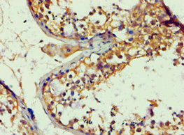
Immunohistochemistry of paraffin-embedded human testis tissue using CSB-PA001890ESR2HU at dilution of 1:100

Immunohistochemistry of paraffin-embedded human testis tissue using CSB-PA001890ESR2HU at dilution of 1:100
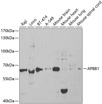
APBB1 Antibody
CSB-PA001890ESR2HU
ApplicationsELISA, ImmunoHistoChemistry
Product group Antibodies
ReactivityHuman
TargetAPBB1
Overview
- SupplierCusabio
- Product NameAPBB1 Antibody
- Delivery Days Customer20
- ApplicationsELISA, ImmunoHistoChemistry
- CertificationResearch Use Only
- ClonalityPolyclonal
- ConjugateUnconjugated
- Gene ID322
- Target nameAPBB1
- Target descriptionamyloid beta precursor protein binding family B member 1
- Target synonymsFE65, MGC:9072, RIR, amyloid beta precursor protein binding family B member 1, adaptor protein FE65a2, amyloid beta (A4) precursor protein-binding, family B, member 1 (Fe65), stat-like protein
- HostRabbit
- IsotypeIgG
- Protein IDO00213
- Protein NameAmyloid beta precursor protein binding family B member 1
- Scientific DescriptionTranscription coregulator that can have both coactivator and corepressor functions. Adapter protein that forms a transcriptionally active complex with the gamma-secretase-derived amyloid precursor protein (APP) intracellular domain. Plays a central role in the response to DNA damage by translocating to the nucleus and inducing apoptosis. May act by specifically recognizing and binding histone H2AX phosphorylated on Tyr-142 (H2AXY142ph) at double-strand breaks (DSBs), recruiting other pro-apoptosis factors such as MAPK8/JNK1. Required for histone H4 acetylation at double-strand breaks (DSBs). Its ability to specifically bind modified histones and chromatin modifying enzymes such as KAT5/TIP60, probably explains its trancription activation activity. Function in association with TSHZ3, SET and HDAC factors as a transcriptional repressor, that inhibits the expression of CASP4. Associates with chromatin in a region surrounding the CASP4 transcriptional start site(s).
- ReactivityHuman
- Storage Instruction-20°C or -80°C
- UNSPSC41116161
Related products
Product group Antibodies
Anti-APBB1 Antibody144-65429
ApplicationsImmunoFluorescence, Western Blot, ImmunoHistoChemistry
ReactivityHuman, Mouse
TargetAPBB1
- SizePrice
Product group Antibodies
Anti-FE65 AntibodyA13762
ApplicationsWestern Blot
ReactivityHuman, Mouse
- SizePrice
Product group Antibodies
FE65 Recombinant Antibody, AbBy Fluor-405 ConjugatedBSM-62200R-BF405
ApplicationsImmunoFluorescence, Western Blot
ReactivityHuman
TargetAPBB1
- SizePrice
Product group Antibodies

Goat anti-APBB1 / FE65EB07373
ApplicationsWestern Blot, ELISA
ReactivityHuman, Mouse
TargetAPBB1
- SizePrice

Product group Antibodies
FE65 antibodyGTX30053
ApplicationsImmunoFluorescence, Western Blot, ImmunoCytoChemistry, ImmunoHistoChemistry, ImmunoHistoChemistry Paraffin
ReactivityHuman, Mouse
TargetAPBB1
- SizePrice
Product group Antibodies

APBB1 / FE65 AntibodyLS-C335192
ApplicationsImmunoFluorescence, Western Blot, ImmunoHistoChemistry
ReactivityHuman, Mouse, Rat
TargetAPBB1
- SizePrice

Product group Antibodies
Anti-APBB1 AntibodyHPA038521
ApplicationsImmunoCytoChemistry, ImmunoHistoChemistry
ReactivityHuman
TargetAPBB1
- SizePrice

Product group Antibodies
Anti-FE65/APBB1 Antibody Picoband(r)PB9684-CARRIER-FREE
ApplicationsWestern Blot, ImmunoHistoChemistry
ReactivityBovine, Human, Mouse, Rat
TargetAPBB1
- SizePrice