


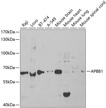
Figure 1. Western blot analysis of FE65 using anti-FE65 antibody (PB9684). Electrophoresis was performed on a 5-20% SDS-PAGE gel at 70V (Stacking gel) / 90V (Resolving gel) for 2-3 hours. Lane 1: Rat Brain Tissue Lysate at 50ug, Lane 2: Mouse Brain Tissue Lysate at 50ug, Lane 3: HELA Whole Cell Lysate at 40ug, Lane 4: U87 Whole Cell Lysate at 40ug. After electrophoresis, proteins were transferred to a nitrocellulose membrane at 150 mA for 50-90 minutes. Blocked the membrane with 5% non-fat milk/TBS for 1.5 hour at RT. The membrane was incubated with rabbit anti-FE65 antigen affinity purified polyclonal antibody (Catalog # PB9684) at 0.5 microg/mL overnight at 4°C, then washed with TBS-0.1%Tween 3 times with 5 minutes each and probed with a goat anti-rabbit IgG-HRP secondary antibody at a dilution of 1:5000 for 1.5 hour at RT. The signal is developed using an Enhanced Chemiluminescent detection (ECL) kit (Catalog # EK1002) with Tanon 5200 system. A specific band was detected for FE65 at approximately 65 kDa. The expected band size for FE65 is at 65 kDa.
. FE65 was detected in a paraffin-embedded section of mouse brain tissue. Heat mediated antigen retrieval was performed in EDTA buffer (pH 8.0, epitope retrieval solution). The tissue section was blocked with 10% goat serum. The tissue section was then incubated with 1 microg/ml rabbit anti-FE65 Antibody (PB9684) overnight at 4°C. Biotinylated goat anti-rabbit IgG was used as secondary antibody and incubated for 30 minutes at 37°C. The tissue section was developed using Strepavidin-Biotin-Complex (SABC) (Catalog # SA1022) with DAB as the chromogen.")
. FE65 was detected in a paraffin-embedded section of rat brain tissue. Heat mediated antigen retrieval was performed in EDTA buffer (pH 8.0, epitope retrieval solution). The tissue section was blocked with 10% goat serum. The tissue section was then incubated with 1 microg/ml rabbit anti-FE65 Antibody (PB9684) overnight at 4°C. Biotinylated goat anti-rabbit IgG was used as secondary antibody and incubated for 30 minutes at 37°C. The tissue section was developed using Strepavidin-Biotin-Complex (SABC) (Catalog # SA1022) with DAB as the chromogen.")
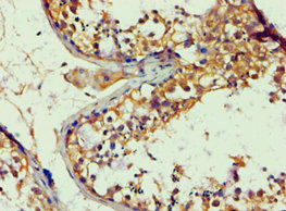
. FE65 was detected in a paraffin-embedded section of human glioma tissue. Heat mediated antigen retrieval was performed in EDTA buffer (pH 8.0, epitope retrieval solution). The tissue section was blocked with 10% goat serum. The tissue section was then incubated with 1 microg/ml rabbit anti-FE65 Antibody (PB9684) overnight at 4°C. Biotinylated goat anti-rabbit IgG was used as secondary antibody and incubated for 30 minutes at 37°C. The tissue section was developed using Strepavidin-Biotin-Complex (SABC) (Catalog # SA1022) with DAB as the chromogen.")
Figure 1. Western blot analysis of FE65 using anti-FE65 antibody (PB9684). Electrophoresis was performed on a 5-20% SDS-PAGE gel at 70V (Stacking gel) / 90V (Resolving gel) for 2-3 hours. Lane 1: Rat Brain Tissue Lysate at 50ug, Lane 2: Mouse Brain Tissue Lysate at 50ug, Lane 3: HELA Whole Cell Lysate at 40ug, Lane 4: U87 Whole Cell Lysate at 40ug. After electrophoresis, proteins were transferred to a nitrocellulose membrane at 150 mA for 50-90 minutes. Blocked the membrane with 5% non-fat milk/TBS for 1.5 hour at RT. The membrane was incubated with rabbit anti-FE65 antigen affinity purified polyclonal antibody (Catalog # PB9684) at 0.5 microg/mL overnight at 4°C, then washed with TBS-0.1%Tween 3 times with 5 minutes each and probed with a goat anti-rabbit IgG-HRP secondary antibody at a dilution of 1:5000 for 1.5 hour at RT. The signal is developed using an Enhanced Chemiluminescent detection (ECL) kit (Catalog # EK1002) with Tanon 5200 system. A specific band was detected for FE65 at approximately 65 kDa. The expected band size for FE65 is at 65 kDa.
Anti-FE65/APBB1 Antibody Picoband(r)
PB9684-CARRIER-FREE
ApplicationsWestern Blot, ImmunoHistoChemistry
Product group Antibodies
ReactivityBovine, Human, Mouse, Rat
TargetAPBB1
Overview
- SupplierBoster Bio
- Product NameAnti-FE65/APBB1 Antibody Picoband(r)
- Delivery Days Customer9
- Application Supplier NoteTested Species: In-house tested species with positive results. By Heat: Boiling the paraffin sections in 10mM citrate buffer, pH6.0, for 20mins is required for the staining of formalin/paraffin sections. Other applications have not been tested. Optimal dilutions should be determined by end users.
- ApplicationsWestern Blot, ImmunoHistoChemistry
- CertificationResearch Use Only
- ClonalityPolyclonal
- Concentration500 ug/ml
- Gene ID322
- Target nameAPBB1
- Target descriptionamyloid beta precursor protein binding family B member 1
- Target synonymsFE65, MGC:9072, RIR, amyloid beta precursor protein binding family B member 1, adaptor protein FE65a2, amyloid beta (A4) precursor protein-binding, family B, member 1 (Fe65), stat-like protein
- HostRabbit
- IsotypeIgG
- Protein IDO00213
- Protein NameAmyloid beta precursor protein binding family B member 1
- Scientific DescriptionBoster Bio Anti-FE65/APBB1 Antibody Picoband® catalog # PB9684. Tested in IHC, WB applications. This antibody reacts with Human, Mouse, Rat. The brand Picoband indicates this is a premium antibody that guarantees superior quality, high affinity, and strong signals with minimal background in Western blot applications. Only our best-performing antibodies are designated as Picoband, ensuring unmatched performance.
- ReactivityBovine, Human, Mouse, Rat
- Storage Instruction-20°C,2°C to 8°C
- UNSPSC12352203
Related products
Product group Antibodies
APBB1 AntibodyCSB-PA001890ESR2HU
ApplicationsELISA, ImmunoHistoChemistry
ReactivityHuman
TargetAPBB1
- SizePrice
Product group Antibodies
Anti-FE65 AntibodyA13762
ApplicationsWestern Blot
ReactivityHuman, Mouse
- SizePrice
Product group Antibodies

Goat anti-APBB1 / FE65EB07373
ApplicationsWestern Blot, ELISA
ReactivityHuman, Mouse
TargetAPBB1
- SizePrice

Product group Antibodies
Anti-APBB1 AntibodyHPA038521
ApplicationsImmunoCytoChemistry, ImmunoHistoChemistry
ReactivityHuman
TargetAPBB1
- SizePrice
Product group Antibodies

APBB1 / FE65 AntibodyLS-C335192
ApplicationsImmunoFluorescence, Western Blot, ImmunoHistoChemistry
ReactivityHuman, Mouse, Rat
TargetAPBB1
- SizePrice

Product group Antibodies
FE65 antibodyGTX30053
ApplicationsImmunoFluorescence, Western Blot, ImmunoCytoChemistry, ImmunoHistoChemistry, ImmunoHistoChemistry Paraffin
ReactivityHuman, Mouse
TargetAPBB1
- SizePrice
Product group Antibodies
FE65 Recombinant Antibody, AbBy Fluor-405 ConjugatedBSM-62200R-BF405
ApplicationsImmunoFluorescence, Western Blot
ReactivityHuman
TargetAPBB1
- SizePrice
Product group Antibodies
Anti-APBB1 Antibody144-65429
ApplicationsImmunoFluorescence, Western Blot, ImmunoHistoChemistry
ReactivityHuman, Mouse
TargetAPBB1
- SizePrice