


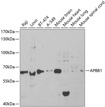
WB analysis of various sample lysates using GTX30053 FE65 antibody. Dilution : 1:1000 Loading : 25μg per lane


WB analysis of various sample lysates using GTX30053 FE65 antibody. Dilution : 1:1000 Loading : 25μg per lane
FE65 antibody
GTX30053
ApplicationsImmunoFluorescence, Western Blot, ImmunoCytoChemistry, ImmunoHistoChemistry, ImmunoHistoChemistry Paraffin
Product group Antibodies
ReactivityHuman, Mouse
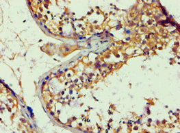
TargetAPBB1
Overview
- SupplierGeneTex
- Product NameFE65 antibody
- Delivery Days Customer7
- Application Supplier NoteWB: 1:50 - 1:200. ICC/IF: 1:50 - 1:200. IHC-P: 1:50 - 1:200. *Optimal dilutions/concentrations should be determined by the researcher.Not tested in other applications.
- ApplicationsImmunoFluorescence, Western Blot, ImmunoCytoChemistry, ImmunoHistoChemistry, ImmunoHistoChemistry Paraffin
- CertificationResearch Use Only
- ClonalityPolyclonal
- ConjugateUnconjugated
- Gene ID322
- Target nameAPBB1
- Target descriptionamyloid beta precursor protein binding family B member 1
- Target synonymsFE65, MGC:9072, RIR, amyloid beta precursor protein binding family B member 1, adaptor protein FE65a2, amyloid beta (A4) precursor protein-binding, family B, member 1 (Fe65), stat-like protein
- HostRabbit
- IsotypeIgG
- Protein IDO00213
- Protein NameAmyloid beta precursor protein binding family B member 1
- Scientific DescriptionThe protein encoded by this gene is a member of the Fe65 protein family. It is an adaptor protein localized in the nucleus. It interacts with the Alzheimers disease amyloid precursor protein (APP), transcription factor CP2/LSF/LBP1 and the low-density lipoprotein receptor-related protein. APP functions as a cytosolic anchoring site that can prevent the gene products nuclear translocation. This encoded protein could play an important role in the pathogenesis of Alzheimers disease. It is thought to regulate transcription. Also it is observed to block cell cycle progression by downregulating thymidylate synthase expression. Multiple alternatively spliced transcript variants encoding different isoforms have been described for this gene. [provided by RefSeq, Mar 2012]
- ReactivityHuman, Mouse
- Storage Instruction-20°C or -80°C,2°C to 8°C
- UNSPSC41116161
Datasheet
Related products
Product group Antibodies
APBB1 AntibodyCSB-PA001890ESR2HU
ApplicationsELISA, ImmunoHistoChemistry
ReactivityHuman
TargetAPBB1
- SizePrice
Product group Antibodies
Anti-FE65 AntibodyA13762
ApplicationsWestern Blot
ReactivityHuman, Mouse
- SizePrice
Product group Antibodies

Goat anti-APBB1 / FE65EB07373
ApplicationsWestern Blot, ELISA
ReactivityHuman, Mouse
TargetAPBB1
- SizePrice

Product group Antibodies
Anti-APBB1 AntibodyHPA038521
ApplicationsImmunoCytoChemistry, ImmunoHistoChemistry
ReactivityHuman
TargetAPBB1
- SizePrice
Product group Antibodies

APBB1 / FE65 AntibodyLS-C335192
ApplicationsImmunoFluorescence, Western Blot, ImmunoHistoChemistry
ReactivityHuman, Mouse, Rat
TargetAPBB1
- SizePrice

Product group Antibodies
Anti-FE65/APBB1 Antibody Picoband(r)PB9684-CARRIER-FREE
ApplicationsWestern Blot, ImmunoHistoChemistry
ReactivityBovine, Human, Mouse, Rat
TargetAPBB1
- SizePrice

Product group Antibodies
FE65 antibody, C-termGTX89278
ApplicationsWestern Blot
ReactivityHuman, Mouse
TargetAPBB1
- SizePrice
Product group Antibodies
FE65 Recombinant Antibody, AbBy Fluor-405 ConjugatedBSM-62200R-BF405
ApplicationsImmunoFluorescence, Western Blot
ReactivityHuman
TargetAPBB1
- SizePrice
Product group Antibodies
Anti-APBB1 Antibody144-65429
ApplicationsImmunoFluorescence, Western Blot, ImmunoHistoChemistry
ReactivityHuman, Mouse
TargetAPBB1
- SizePrice