

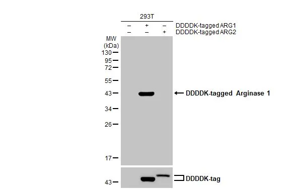
Non-transfected (–) and transfected (+) 293T whole cell extracts (30 μg) were separated by 12% SDS-PAGE, and the membrane was blotted with Arginase 1 antibody [HL1891] (GTX637640) diluted at 1:2500. The HRP-conjugated anti-rabbit IgG antibody (GTX213110-01) was used to detect the primary antibody.
![Various tissue extracts (50 μg) were separated by 12% SDS-PAGE, and the membrane was blotted with Arginase 1 antibody [HL1891] (GTX637640) diluted at 1:500. The HRP-conjugated anti-rabbit IgG antibody (GTX213110-01) was used to detect the primary antibody.](https://www.genetex.com/upload/website/prouct_img/normal/GTX637640/GTX637640_T-44837_20230203_WB_M_tissue_23020621_275.webp "Various tissue extracts (50 μg) were separated by 12% SDS-PAGE, and the membrane was blotted with Arginase 1 antibody [HL1891] (GTX637640) diluted at 1:500. The HRP-conjugated anti-rabbit IgG antibody (GTX213110-01) was used to detect the primary antibody.")
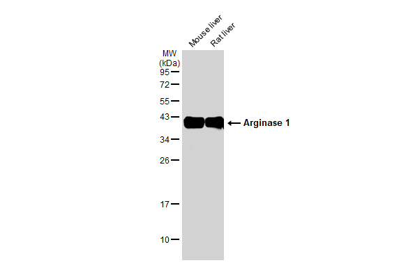
![Rat tissue extract (5 μg) was separated by 12% SDS-PAGE, and the membrane was blotted with Arginase 1 antibody [HL1891] (GTX637640) diluted at 1:40000. The HRP-conjugated anti-rabbit IgG antibody (GTX213110-01) was used to detect the primary antibody.](https://www.genetex.com/upload/website/prouct_img/normal/GTX637640/GTX637640_T-44837_20230317_WB_R_liver_23032022_533.webp "Rat tissue extract (5 μg) was separated by 12% SDS-PAGE, and the membrane was blotted with Arginase 1 antibody [HL1891] (GTX637640) diluted at 1:40000. The HRP-conjugated anti-rabbit IgG antibody (GTX213110-01) was used to detect the primary antibody.")
![Human tissue extract (5 μg) was separated by 12% SDS-PAGE, and the membrane was blotted with Arginase 1 antibody [HL1891] (GTX637640) diluted at 1:20000. The HRP-conjugated anti-rabbit IgG antibody (GTX213110-01) was used to detect the primary antibody.](https://www.genetex.com/upload/website/prouct_img/normal/GTX637640/GTX637640_T-44837_20230317_WB_liver_23032022_165.webp "Human tissue extract (5 μg) was separated by 12% SDS-PAGE, and the membrane was blotted with Arginase 1 antibody [HL1891] (GTX637640) diluted at 1:20000. The HRP-conjugated anti-rabbit IgG antibody (GTX213110-01) was used to detect the primary antibody.")
![Arginase 1 antibody [HL1891] detects Arginase 1 protein at cytoplasm by immunohistochemical analysis. Sample: Paraffin-embedded rat liver. Arginase 1 stained by Arginase 1 antibody [HL1891] (GTX637640) diluted at 1:100. Antigen Retrieval: Citrate buffer, pH 6.0, 15 min](https://www.genetex.com/upload/website/prouct_img/normal/GTX637640/GTX637640_T-44837_20230331_IHC-P_R_23032819_778.webp "Arginase 1 antibody [HL1891] detects Arginase 1 protein at cytoplasm by immunohistochemical analysis. Sample: Paraffin-embedded rat liver. Arginase 1 stained by Arginase 1 antibody [HL1891] (GTX637640) diluted at 1:100. Antigen Retrieval: Citrate buffer, pH 6.0, 15 min")
![Arginase 1 antibody [HL1891] detects Arginase 1 protein at cytoplasm by immunohistochemical analysis. Sample: Paraffin-embedded mouse liver. Arginase 1 stained by Arginase 1 antibody [HL1891] (GTX637640) diluted at 1:100. Antigen Retrieval: Citrate buffer, pH 6.0, 15 min](https://www.genetex.com/upload/website/prouct_img/normal/GTX637640/GTX637640_T-44837_20230331_IHC-P_M_23032819_779.webp "Arginase 1 antibody [HL1891] detects Arginase 1 protein at cytoplasm by immunohistochemical analysis. Sample: Paraffin-embedded mouse liver. Arginase 1 stained by Arginase 1 antibody [HL1891] (GTX637640) diluted at 1:100. Antigen Retrieval: Citrate buffer, pH 6.0, 15 min")
![Non-transfected (–) and transfected (+) HepG2 whole cell extracts (30 μg) were separated by 12% SDS-PAGE, and the membrane was blotted with Arginase 1 antibody [HL1891] (GTX637640) diluted at 1:500. The HRP-conjugated anti-rabbit IgG antibody (GTX213110-01) was used to detect the primary antibody, and the signal was developed with Trident ECL plus-Enhanced.](https://www.genetex.com/upload/website/prouct_img/normal/GTX637640/GTX637640_T-44837_20230630_WB_shRNA_watermark_23070401_549.webp "Non-transfected (–) and transfected (+) HepG2 whole cell extracts (30 μg) were separated by 12% SDS-PAGE, and the membrane was blotted with Arginase 1 antibody [HL1891] (GTX637640) diluted at 1:500. The HRP-conjugated anti-rabbit IgG antibody (GTX213110-01) was used to detect the primary antibody, and the signal was developed with Trident ECL plus-Enhanced.")
Non-transfected (–) and transfected (+) 293T whole cell extracts (30 μg) were separated by 12% SDS-PAGE, and the membrane was blotted with Arginase 1 antibody [HL1891] (GTX637640) diluted at 1:2500. The HRP-conjugated anti-rabbit IgG antibody (GTX213110-01) was used to detect the primary antibody.
Arginase 1 antibody [HL1891]
GTX637640
ApplicationsWestern Blot, ImmunoHistoChemistry, ImmunoHistoChemistry Paraffin
Product group Antibodies
ReactivityHuman, Mouse, Rat
TargetARG1
Overview
- SupplierGeneTex
- Product NameArginase 1 antibody [HL1891]
- Delivery Days Customer9
- Application Supplier NoteWB: 1:500-1:3000. *Optimal dilutions/concentrations should be determined by the researcher.Not tested in other applications.
- ApplicationsWestern Blot, ImmunoHistoChemistry, ImmunoHistoChemistry Paraffin
- CertificationResearch Use Only
- ClonalityMonoclonal
- Clone IDHL1891
- Concentration2 mg/ml
- ConjugateUnconjugated
- Gene ID383
- Target nameARG1
- Target descriptionarginase 1
- Target synonymsarginase-1, arginase, liver, liver-type arginase, type I arginase
- HostRabbit
- IsotypeIgG
- Protein IDP05089
- Protein NameArginase-1
- Scientific DescriptionArginase catalyzes the hydrolysis of arginine to ornithine and urea. At least two isoforms of mammalian arginase exist (types I and II) which differ in their tissue distribution, subcellular localization, immunologic crossreactivity and physiologic function. The type I isoform encoded by this gene, is a cytosolic enzyme and expressed predominantly in the liver as a component of the urea cycle. Inherited deficiency of this enzyme results in argininemia, an autosomal recessive disorder characterized by hyperammonemia. Two transcript variants encoding different isoforms have been found for this gene. [provided by RefSeq, Sep 2011]
- ReactivityHuman, Mouse, Rat
- Storage Instruction-20°C or -80°C,2°C to 8°C
- UNSPSC12352203
Datasheet
Related products
Product group Antibodies

Anti-Arginase 1 [mAb5]Ab03081-1.1
ApplicationsElectron Microscopy, Neutralisation/Blocking
ReactivityHuman
TargetARG1
- SizePrice

Product group Antibodies
Anti-ARG1 Antibody144-01847
ApplicationsWestern Blot, ImmunoHistoChemistry
ReactivityHuman, Mouse, Rat
TargetARG1
- SizePrice

Product group Antibodies
Anti-ARG1 AntibodyAMAB90545
ApplicationsWestern Blot, ImmunoHistoChemistry
ReactivityHuman
TargetARG1
- SizePrice

Product group Antibodies
Anti-liver Arginase/ARG1 Antibody Picoband(r)A01106-CARRIER-FREE
ApplicationsWestern Blot, ELISA, ImmunoHistoChemistry
ReactivityHuman, Monkey, Mouse, Rat
TargetARG1
- SizePrice
![IHC-P analysis of human liver tissue using GTX04426 Arginase 1 antibody [MSVA-511R] HistoMAX?. Strong nuclear and cytoplasmic arginase 1 expression in all hepatocytes in a normal liver.](https://www.genetex.com/upload/website/prouct_img/normal/GTX04426/GTX04426_20230728_IHC-P_4_23072722_512.webp)
Product group Antibodies
ApplicationsImmunoHistoChemistry, ImmunoHistoChemistry Paraffin
ReactivityHuman
TargetARG1
- SizePrice
Product group Antibodies
References
Arginase 1 antibodyGTX109242
ApplicationsFlow Cytometry, ImmunoFluorescence, ImmunoPrecipitation, Western Blot, ELISA, ImmunoCytoChemistry, ImmunoHistoChemistry, ImmunoHistoChemistry Frozen, ImmunoHistoChemistry Paraffin
ReactivityHuman, Mouse, Rat
TargetARG1
- SizePrice
Product group Antibodies
References
Arginase 1 antibodyGTX113131
ApplicationsImmunoFluorescence, Western Blot, ELISA, ImmunoCytoChemistry, ImmunoHistoChemistry, ImmunoHistoChemistry Paraffin
ReactivityHuman, Mouse, Rat
TargetARG1
- SizePrice
Product group Antibodies
References
Arginase 1 antibody [GT5811]GTX634218
ApplicationsFlow Cytometry, Western Blot, ELISA, ImmunoHistoChemistry, ImmunoHistoChemistry Paraffin
ReactivityHuman, Mouse, Rat
TargetARG1
- SizePrice
Product group Antibodies
ARG1 Polyclonal AntibodyCAC14814
ApplicationsWestern Blot, ELISA, ImmunoHistoChemistry
ReactivityMouse, Rat
TargetARG1
- SizePrice