

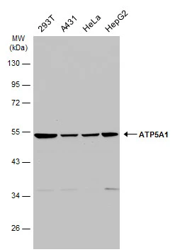
Various whole cell extracts (30 μg) were separated by 10% SDS-PAGE, and the membrane was blotted with ATP5A1 antibody (GTX101741) diluted at 1:2000. The HRP-conjugated anti-rabbit IgG antibody (GTX213110-01) was used to detect the primary antibody.
![ATP5A1 antibody [C2C3], C-term detects ATP5A1 protein at mitochondria by immunofluorescent analysis. Sample: HeLa cells were fixed in 4% paraformaldehyde at RT for 15 min. Green: ATP5A1 protein stained by ATP5A1 antibody [C2C3], C-term (GTX101741) diluted at 1:500. Blue: Hoechst 33342 staining. Scale bar = 10 μm.](https://www.genetex.com/upload/website/prouct_img/normal/GTX101741/GTX101741_41871_20170524_IFA_w_23060100_945.webp "ATP5A1 antibody [C2C3], C-term detects ATP5A1 protein at mitochondria by immunofluorescent analysis. Sample: HeLa cells were fixed in 4% paraformaldehyde at RT for 15 min. Green: ATP5A1 protein stained by ATP5A1 antibody [C2C3], C-term (GTX101741) diluted at 1:500. Blue: Hoechst 33342 staining. Scale bar = 10 μm.")
![HepG2 and mitochondria extracts (30 μg) were separated by SDS-PAGE, and the membrane was blotted with ATP5A1 antibody [C2C3], C-term (GTX101741) diluted at 1:2000. The HRP-conjugated anti-rabbit IgG antibody (GTX213110-01) was used to detect the primary antibody.](https://www.genetex.com/upload/website/prouct_img/normal/GTX101741/GTX101741_41871_20191129_WB_Fraction_w_23060100_455.webp "HepG2 and mitochondria extracts (30 μg) were separated by SDS-PAGE, and the membrane was blotted with ATP5A1 antibody [C2C3], C-term (GTX101741) diluted at 1:2000. The HRP-conjugated anti-rabbit IgG antibody (GTX213110-01) was used to detect the primary antibody.")
![ATP5A1 antibody [C2C3], C-term detects ATP5A1 protein at cytosol on human colon carcinoma by immunohistochemical analysis. Sample: Paraffin-embedded colon carcinoma. ATP5A1 antibody [C2C3], C-term (GTX101741) dilution: 1:200.
Antigen Retrieval: Trilogy? (EDTA based, pH 8.0) buffer, 15min](https://www.genetex.com/upload/website/prouct_img/normal/GTX101741/GTX101741_40128_IHC_w_23060100_595.webp "ATP5A1 antibody [C2C3], C-term detects ATP5A1 protein at cytosol on human colon carcinoma by immunohistochemical analysis. Sample: Paraffin-embedded colon carcinoma. ATP5A1 antibody [C2C3], C-term (GTX101741) dilution: 1:200.
Antigen Retrieval: Trilogy? (EDTA based, pH 8.0) buffer, 15min")
![Various tissue extracts (50 μg) were separated by 10% SDS-PAGE, and the membrane was blotted with ATP5A1 antibody [C2C3], C-term (GTX101741) diluted at 1:1000.](https://www.genetex.com/upload/website/prouct_img/normal/GTX101741/GTX101741_41871_20160818_WB_M_R_w_23060100_956.webp "Various tissue extracts (50 μg) were separated by 10% SDS-PAGE, and the membrane was blotted with ATP5A1 antibody [C2C3], C-term (GTX101741) diluted at 1:1000.")
![ATP5A1 antibody [C2C3], C-term detects ATP5A1 protein at cytoplasm on mouse stomach by immunohistochemical analysis. Sample: Paraffin-embedded mouse stomach. ATP5A1 antibody [C2C3], C-term (GTX101741) dilution: 1:500.
Antigen Retrieval: Trilogy? (EDTA based, pH 8.0) buffer, 15min](https://www.genetex.com/upload/website/prouct_img/normal/GTX101741/GTX101741_40128_IHC_M_w_23060100_870.webp "ATP5A1 antibody [C2C3], C-term detects ATP5A1 protein at cytoplasm on mouse stomach by immunohistochemical analysis. Sample: Paraffin-embedded mouse stomach. ATP5A1 antibody [C2C3], C-term (GTX101741) dilution: 1:500.
Antigen Retrieval: Trilogy? (EDTA based, pH 8.0) buffer, 15min")
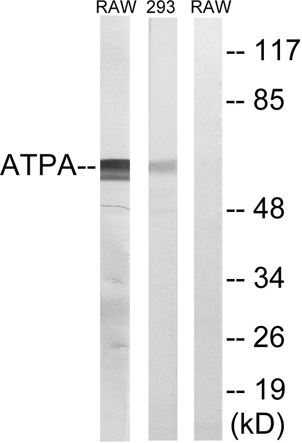
 were separated by 10% SDS-PAGE, and the membrane was blotted with ATP5A1 antibody (GTX101741) diluted by 1:2000.")
![Non-transfected (–) and transfected (+) 293T whole cell extracts (30 μg) were separated by 10% SDS-PAGE, and the membrane was blotted with ATP5A1 antibody [C2C3], C-term (GTX101741) diluted at 1:1000. The HRP-conjugated anti-rabbit IgG antibody (GTX213110-01) was used to detect the primary antibody.](https://www.genetex.com/upload/website/prouct_img/normal/GTX101741/GTX101741_41871_20180831_WB_B_w_23060100_199.webp "Non-transfected (–) and transfected (+) 293T whole cell extracts (30 μg) were separated by 10% SDS-PAGE, and the membrane was blotted with ATP5A1 antibody [C2C3], C-term (GTX101741) diluted at 1:1000. The HRP-conjugated anti-rabbit IgG antibody (GTX213110-01) was used to detect the primary antibody.")
![ATP5A1 antibody [C2C3], C-term detects ATP5A1 protein by immunohistochemical analysis. Sample: Frozen sectioned adult mouse retina. Green: ATP5A1 protein stained by ATP5A1 antibody [C2C3], C-term (GTX101741) diluted at 1:250. Red: Protein kinase C alpha staining. Blue: Fluoroshield with DAPI (GTX30920).](https://www.genetex.com/upload/website/prouct_img/normal/GTX101741/GTX101741_41871_20160808_IHC-Fr_w_23060100_186.webp "ATP5A1 antibody [C2C3], C-term detects ATP5A1 protein by immunohistochemical analysis. Sample: Frozen sectioned adult mouse retina. Green: ATP5A1 protein stained by ATP5A1 antibody [C2C3], C-term (GTX101741) diluted at 1:250. Red: Protein kinase C alpha staining. Blue: Fluoroshield with DAPI (GTX30920).")
![ATP5A1 antibody [C2C3], C-term detects ATP5A1 protein at membrane on mouse lung by immunohistochemical analysis. Sample: Paraffin-embedded mouse lung. ATP5A1 antibody [C2C3], C-term (GTX101741) dilution: 1:500.
Antigen Retrieval: Trilogy? (EDTA based, pH 8.0) buffer, 15min](https://www.genetex.com/upload/website/prouct_img/normal/GTX101741/GTX101741_40128_IHC_M_2_w_23060100_970.webp "ATP5A1 antibody [C2C3], C-term detects ATP5A1 protein at membrane on mouse lung by immunohistochemical analysis. Sample: Paraffin-embedded mouse lung. ATP5A1 antibody [C2C3], C-term (GTX101741) dilution: 1:500.
Antigen Retrieval: Trilogy? (EDTA based, pH 8.0) buffer, 15min")
Various whole cell extracts (30 μg) were separated by 10% SDS-PAGE, and the membrane was blotted with ATP5A1 antibody (GTX101741) diluted at 1:2000. The HRP-conjugated anti-rabbit IgG antibody (GTX213110-01) was used to detect the primary antibody.
ATP5A1 antibody [C2C3], C-term
GTX101741
ApplicationsImmunoFluorescence, Western Blot, ImmunoCytoChemistry, ImmunoHistoChemistry, ImmunoHistoChemistry Frozen, ImmunoHistoChemistry Paraffin
Product group Antibodies
ReactivityHuman, Mouse, Rat
TargetATP5F1A
Overview
- SupplierGeneTex
- Product NameATP5A1 antibody [C2C3], C-term
- Delivery Days Customer9
- Application Supplier NoteWB: 1:500-1:3000. ICC/IF: 1:100-1:1000. IHC-P: 1:100-1:1000. IHC-Fr: 1:100-1:1000. *Optimal dilutions/concentrations should be determined by the researcher.Not tested in other applications.
- ApplicationsImmunoFluorescence, Western Blot, ImmunoCytoChemistry, ImmunoHistoChemistry, ImmunoHistoChemistry Frozen, ImmunoHistoChemistry Paraffin
- CertificationResearch Use Only
- ClonalityPolyclonal
- Concentration0.66 mg/ml
- ConjugateUnconjugated
- Gene ID498
- Target nameATP5F1A
- Target descriptionATP synthase F1 subunit alpha
- Target synonymsATP5A, ATP5A1, ATP5AL2, ATPM, COXPD22, HEL-S-123m, MC5DN4, MC5DN4A, MC5DN4B, MOM2, OMR, ORM, hATP1, ATP synthase F(1) complex subunit alpha, mitochondrial, ATP synthase alpha chain, mitochondrial, ATP synthase, H+ transporting, mitochondrial F1 complex, alpha subunit 1, cardiac muscle, ATP sythase (F1-ATPase) alpha subunit, epididymis secretory sperm binding protein Li 123m, mitochondrial ATP synthetase, oligomycin-resistant
- HostRabbit
- IsotypeIgG
- Protein IDP25705
- Protein NameATP synthase F(1) complex subunit alpha, mitochondrial
- Scientific DescriptionThis gene encodes a subunit of mitochondrial ATP synthase. Mitochondrial ATP synthase catalyzes ATP synthesis, using an electrochemical gradient of protons across the inner membrane during oxidative phosphorylation. ATP synthase is composed of two linked multi-subunit complexes: the soluble catalytic core, F1, and the membrane-spanning component, Fo, comprising the proton channel. The catalytic portion of mitochondrial ATP synthase consists of 5 different subunits (alpha, beta, gamma, delta, and epsilon) assembled with a stoichiometry of 3 alpha, 3 beta, and a single representative of the other 3. The proton channel consists of three main subunits (a, b, c). This gene encodes the alpha subunit of the catalytic core. Alternatively spliced transcript variants encoding the same protein have been identified. Pseudogenes of this gene are located on chromosomes 9, 2, and 16. [provided by RefSeq]
- ReactivityHuman, Mouse, Rat
- Storage Instruction-20°C or -80°C,2°C to 8°C
- UNSPSC41116161
Datasheet
Related products
Product group Antibodies
Anti-ATP5A1 AntibodyA96952
ApplicationsWestern Blot, ELISA
ReactivityHuman, Mouse, Rat
- SizePrice
Product group Antibodies
Anti-ATP5A1 Antibody144-05884
ApplicationsImmunoFluorescence, ImmunoPrecipitation, Western Blot, ImmunoHistoChemistry
ReactivityHuman, Mouse, Rat
TargetATP5F1A
- SizePrice

Product group Antibodies
Anti-ATP5F1A Antibody Picoband(r)A32267-2-CARRIER-FREE
ApplicationsFlow Cytometry, ImmunoFluorescence, Western Blot, ELISA, ImmunoCytoChemistry, ImmunoHistoChemistry
ReactivityHuman, Mouse, Rat
TargetATP5F1A
- SizePrice
Product group Antibodies
ATP5A Recombinant Antibody, Biotin ConjugatedBSM-61444R-BIOTIN
ApplicationsWestern Blot, ImmunoHistoChemistry, ImmunoHistoChemistry Frozen, ImmunoHistoChemistry Paraffin
ReactivityHuman, Mouse, Rat
TargetATP5F1A
- SizePrice

Product group Antibodies
ATP5A1 AntibodyCSB-PA000951
ApplicationsWestern Blot, ELISA, ImmunoHistoChemistry
ReactivityHuman, Mouse, Rat
TargetATP5F1A
- SizePrice
Product group Antibodies
References

Goat anti-ATP5A1EB12389
ApplicationsWestern Blot, ELISA
ReactivityBovine, Canine, Human, Mouse, Porcine, Rat
TargetATP5F1A
- SizePrice
Product group Antibodies
Atp5F1A Polyclonal AntibodyCAC07482
ApplicationsWestern Blot, ELISA
TargetATP5F1A
- SizePrice
Product group Antibodies

ApplicationsWestern Blot, ELISA
ReactivityHuman
TargetATP5F1A
- SizePrice

Product group Antibodies
Anti-ATP5A1 AntibodyHPA040622
ApplicationsWestern Blot, ImmunoHistoChemistry
ReactivityHuman
TargetATP5F1A
- SizePrice