

Various whole cell extracts (30 μg) were separated by 5% SDS-PAGE, and the membrane was blotted with ATRX antibody [C3], C-term (GTX101310) diluted at 1:1000. The HRP-conjugated anti-rabbit IgG antibody (GTX213110-01) was used to detect the primary antibody.
![Rad54 antibody [C3], C-term detects Rad54 protein at nucleus on human endometrial carcinoma by immunohistochemical analysis. Sample: Paraffin-embedded human endometrial carcinoma. Rad54 antibody [C3], C-term (GTX101310) dilution: 1:500.](https://www.genetex.com/upload/website/prouct_img/normal/GTX101310/GTX101310_41633_IHC_3_w_23060100_973.webp "Rad54 antibody [C3], C-term detects Rad54 protein at nucleus on human endometrial carcinoma by immunohistochemical analysis. Sample: Paraffin-embedded human endometrial carcinoma. Rad54 antibody [C3], C-term (GTX101310) dilution: 1:500.")
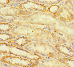
![ATRX antibody [C3], C-term detects ATRX protein at cytoplasm and nucleus by immunohistochemical analysis. Sample: Paraffin-embedded human lung cancer. ATRX stained by ATRX antibody [C3], C-term (GTX101310) diluted at 1:500. Antigen Retrieval: Citrate buffer, pH 6.0, 15 min](https://www.genetex.com/upload/website/prouct_img/normal/GTX101310/GTX101310_43635_20190830_IHC-P_w_23060100_505.webp "ATRX antibody [C3], C-term detects ATRX protein at cytoplasm and nucleus by immunohistochemical analysis. Sample: Paraffin-embedded human lung cancer. ATRX stained by ATRX antibody [C3], C-term (GTX101310) diluted at 1:500. Antigen Retrieval: Citrate buffer, pH 6.0, 15 min")
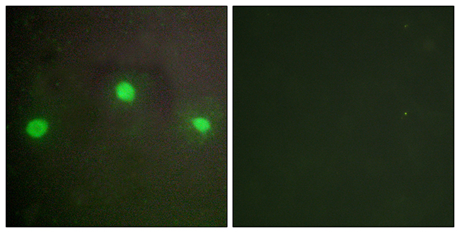
![ATRX antibody [C3], C-term detects ATRX protein at nucleus by immunofluorescent analysis. Sample: HeLa cells were fixed in 4% paraformaldehyde at RT for 15 min. Green: ATRX stained by ATRX antibody [C3], C-term (GTX101310) diluted at 1:1000. Red: phalloidin, a cytoskeleton marker, diluted at 1:200. Scale bar= 10 μm.](https://www.genetex.com/upload/website/prouct_img/normal/GTX101310/GTX101310_43635_20200311_ICC_IF_w_23060100_827.webp "ATRX antibody [C3], C-term detects ATRX protein at nucleus by immunofluorescent analysis. Sample: HeLa cells were fixed in 4% paraformaldehyde at RT for 15 min. Green: ATRX stained by ATRX antibody [C3], C-term (GTX101310) diluted at 1:1000. Red: phalloidin, a cytoskeleton marker, diluted at 1:200. Scale bar= 10 μm.")
![Non-transfected (–) and transfected (+) 293T whole cell extracts (30 μg) were separated by 5% SDS-PAGE, and the membrane was blotted with ATRX antibody [C3], C-term (GTX101310) diluted at 1:2000. The HRP-conjugated anti-rabbit IgG antibody (GTX213110-01) was used to detect the primary antibody.](https://www.genetex.com/upload/website/prouct_img/normal/GTX101310/GTX101310_42165_20161110_WB_shRNA_watermark_w_23060100_970.webp "Non-transfected (–) and transfected (+) 293T whole cell extracts (30 μg) were separated by 5% SDS-PAGE, and the membrane was blotted with ATRX antibody [C3], C-term (GTX101310) diluted at 1:2000. The HRP-conjugated anti-rabbit IgG antibody (GTX213110-01) was used to detect the primary antibody.")
![Immunoprecipitation of ATRX protein from 293T whole cell extracts using 5 μg of ATRX antibody [C3], C-term (GTX101310). Western blot analysis was performed using ATRX antibody [C3], C-term (GTX101310). EasyBlot anti-Rabbit IgG (GTX221666-01) was used as a secondary reagent.](https://www.genetex.com/upload/website/prouct_img/normal/GTX101310/GTX101310_41787_20150209_IP_w_23060100_755.webp "Immunoprecipitation of ATRX protein from 293T whole cell extracts using 5 μg of ATRX antibody [C3], C-term (GTX101310). Western blot analysis was performed using ATRX antibody [C3], C-term (GTX101310). EasyBlot anti-Rabbit IgG (GTX221666-01) was used as a secondary reagent.")
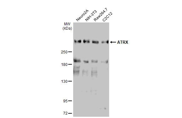
![Various whole cell extracts (30 μg) were separated by 5% SDS-PAGE, and the membrane was blotted with ATRX antibody [C3], C-term (GTX101310) diluted at 1:1000. The HRP-conjugated anti-rabbit IgG antibody (GTX213110-01) was used to detect the primary antibody.Corresponding RNA expression data for the same cell lines are based on Human Protein Atlas program.](https://www.genetex.com/upload/website/prouct_img/normal/GTX101310/GTX101310_43341_20190524_WB_TPM_watremark_w_23060100_381.webp "Various whole cell extracts (30 μg) were separated by 5% SDS-PAGE, and the membrane was blotted with ATRX antibody [C3], C-term (GTX101310) diluted at 1:1000. The HRP-conjugated anti-rabbit IgG antibody (GTX213110-01) was used to detect the primary antibody.Corresponding RNA expression data for the same cell lines are based on Human Protein Atlas program.")
![ATRX antibody [C3], C-term detects ATRX protein at cytoplasm and nucleus by immunohistochemical analysis. Sample: Paraffin-embedded human lung cancer. ATRX stained by ATRX antibody [C3], C-term (GTX101310) diluted at 1:2500. Antigen Retrieval: Citrate buffer, pH 6.0, 15 min](https://www.genetex.com/upload/website/prouct_img/normal/GTX101310/GTX101310_44426_20211015_IHC-P_w_23060100_829.webp "ATRX antibody [C3], C-term detects ATRX protein at cytoplasm and nucleus by immunohistochemical analysis. Sample: Paraffin-embedded human lung cancer. ATRX stained by ATRX antibody [C3], C-term (GTX101310) diluted at 1:2500. Antigen Retrieval: Citrate buffer, pH 6.0, 15 min")
![ATRX antibody [C3], C-term detects ATRX protein at nucleus by immunohistochemical analysis. Sample: Paraffin-embedded human breast carcinoma. ATRX stained by ATRX antibody [C3], C-term (GTX101310) diluted at 1:2000.
Antigen Retrieval: Citrate buffer, pH 6.0, 15 min](https://www.genetex.com/upload/website/prouct_img/normal/GTX101310/GTX101310_43005_20180213_IHC-P_w_23060100_646.webp "ATRX antibody [C3], C-term detects ATRX protein at nucleus by immunohistochemical analysis. Sample: Paraffin-embedded human breast carcinoma. ATRX stained by ATRX antibody [C3], C-term (GTX101310) diluted at 1:2000.
Antigen Retrieval: Citrate buffer, pH 6.0, 15 min")
![Various whole cell extracts (30 μg) were separated by 5% SDS-PAGE, and the membrane was blotted with ATRX antibody [C3], C-term (GTX101310) diluted at 1:1000. The HRP-conjugated anti-rabbit IgG antibody (GTX213110-01) was used to detect the primary antibody.](https://www.genetex.com/upload/website/prouct_img/normal/GTX101310/GTX101310_44426_20210910_WB_23122619_597.webp "Various whole cell extracts (30 μg) were separated by 5% SDS-PAGE, and the membrane was blotted with ATRX antibody [C3], C-term (GTX101310) diluted at 1:1000. The HRP-conjugated anti-rabbit IgG antibody (GTX213110-01) was used to detect the primary antibody.")
Various whole cell extracts (30 μg) were separated by 5% SDS-PAGE, and the membrane was blotted with ATRX antibody [C3], C-term (GTX101310) diluted at 1:1000. The HRP-conjugated anti-rabbit IgG antibody (GTX213110-01) was used to detect the primary antibody.
ATRX antibody [C3], C-term
GTX101310
ApplicationsImmunoFluorescence, ImmunoPrecipitation, Western Blot, ImmunoCytoChemistry, ImmunoHistoChemistry, ImmunoHistoChemistry Paraffin
Product group Antibodies
ReactivityHuman, Mouse, Rat
TargetATRX
Overview
- SupplierGeneTex
- Product NameATRX antibody [C3], C-term
- Delivery Days Customer9
- Application Supplier NoteWB: 1:500-1:3000. ICC/IF: 1:100-1:1000. IHC-P: 1:100-1:1000. IP: 1:100-1:500. *Optimal dilutions/concentrations should be determined by the researcher.Not tested in other applications.
- ApplicationsImmunoFluorescence, ImmunoPrecipitation, Western Blot, ImmunoCytoChemistry, ImmunoHistoChemistry, ImmunoHistoChemistry Paraffin
- CertificationResearch Use Only
- ClonalityPolyclonal
- Concentration0.27 mg/ml
- ConjugateUnconjugated
- Gene ID546
- Target nameATRX
- Target descriptionATRX chromatin remodeler
- Target synonymsJMS, MRX52, RAD54, RAD54L, XH2, XNP, ZNF-HX, transcriptional regulator ATRX, ATP-dependent helicase ATRX, X-linked helicase II, X-linked nuclear protein
- HostRabbit
- IsotypeIgG
- Protein IDP46100
- Protein NameTranscriptional regulator ATRX
- Scientific DescriptionThe protein encoded by this gene contains an ATPase/helicase domain, and thus it belongs to the SWI/SNF family of chromatin remodeling proteins. The mutations of this gene are associated with an X-linked mental retardation (XLMR) syndrome most often accompanied by alpha-thalassemia (ATRX) syndrome. These mutations have been shown to cause diverse changes in the pattern of DNA methylation, which may provide a link between chromatin remodeling, DNA methylation, and gene expression in developmental processes. This protein is found to undergo cell cycle-dependent phosphorylation, which regulates its nuclear matrix and chromatin association, and suggests its involvement in the gene regulation at interphase and chromosomal segregation in mitosis. Multiple alternatively spliced transcript variants encoding distinct isoforms have been reported. [provided by RefSeq]
- ReactivityHuman, Mouse, Rat
- Storage Instruction-20°C or -80°C,2°C to 8°C
- UNSPSC41116161
Datasheet
Related products
Product group Antibodies
Anti-ATRX AntibodyA99468
ApplicationsImmunoFluorescence, ELISA
ReactivityHuman, Mouse
- SizePrice
Product group Antibodies

Anti-ATRX [20A2]Ab03305-10.0
ApplicationsWestern Blot, ELISA, ImmunoHistoChemistry
ReactivityHuman
TargetATRX
- SizePrice
Product group Antibodies
Anti-ATRX Antibody101-10135
ApplicationsWestern Blot, ELISA
TargetATRX
- SizePrice

Product group Antibodies
Anti-ATRX AntibodyAMAB90784
ApplicationsWestern Blot, ImmunoCytoChemistry, ImmunoHistoChemistry
ReactivityHuman
TargetATRX
- SizePrice
Product group Antibodies

ATRX AntibodyLS-C831157
ApplicationsELISA, ImmunoHistoChemistry
ReactivityHuman, Mouse
TargetATRX
- SizePrice

Product group Antibodies
Anti-ATRX Antibody Picoband(r)A00203-2-CARRIER-FREE
ApplicationsFlow Cytometry, Western Blot, ELISA, ImmunoHistoChemistry
ReactivityHuman, Mouse, Rat
TargetATRX
- SizePrice
Product group Antibodies
ATRX Polyclonal AntibodyBS-24302R
ApplicationsWestern Blot, ELISA
ReactivityBovine, Canine, Chicken, Equine, Human, Mouse, Porcine, Rabbit, Rat, Sheep
TargetATRX
- SizePrice
Product group Antibodies
ATRX AntibodyCSB-PA002437LA01HU
ApplicationsImmunoFluorescence, ELISA, ImmunoHistoChemistry
ReactivityHuman
TargetATRX
- SizePrice
![IHC-P analysis of human prostate tissue section using GTX02592 ATRX antibody [rATRX/3446].](https://www.genetex.com/upload/website/prouct_img/normal/GTX02592/GTX02592_20210319_IHC-P_w_23053122_951.webp)
Product group Antibodies
ATRX antibody [rATRX/3446]GTX02592
ApplicationsImmunoHistoChemistry, ImmunoHistoChemistry Paraffin
ReactivityHuman
TargetATRX
- SizePrice
![IHC-P analysis of human prostate carcinoma section using GTX02593 ATRX antibody [ATRX/2900R].](https://www.genetex.com/upload/website/prouct_img/normal/GTX02593/GTX02593_20210319_IHC-P_w_23053122_524.webp)
Product group Antibodies
ATRX antibody [ATRX/2900R]GTX02593
ApplicationsImmunoHistoChemistry, ImmunoHistoChemistry Paraffin
ReactivityHuman
TargetATRX
- SizePrice