

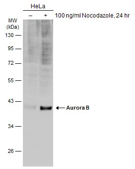
Untreated (–) and treated (+) HeLa whole cell extracts (30 μg) were separated by 10% SDS-PAGE, and the membrane was blotted with Aurora B antibody (GTX132702) diluted at 1:1000. The HRP-conjugated anti-rabbit IgG antibody (GTX213110-01) was used to detect the primary antibody.
 and transfected (+) 293T whole cell extracts (30 μg) were separated by 10% SDS-PAGE, and the membrane was blotted with Aurora B antibody (GTX132702) diluted at 1:5000. The HRP-conjugated anti-rabbit IgG antibody (GTX213110-01) was used to detect the primary antibody.")
![Aurora B antibody detects Aurora B protein by immunofluorescent analysis. Sample: HeLa cells were fixed in 4% paraformaldehyde at RT for 15 min. Green: Aurora B stained by Aurora B antibody (GTX132702) diluted at 1:500. Red: alpha Tubulin, stained by alpha Tubulin antibody [GT114] (GTX628802) diluted at 1:500. Blue: Hoechst 33342 staining.](https://www.genetex.com/upload/website/prouct_img/normal/GTX132702/GTX132702_43173_20181205_ICC_IF_w_23060523_481.webp "Aurora B antibody detects Aurora B protein by immunofluorescent analysis. Sample: HeLa cells were fixed in 4% paraformaldehyde at RT for 15 min. Green: Aurora B stained by Aurora B antibody (GTX132702) diluted at 1:500. Red: alpha Tubulin, stained by alpha Tubulin antibody [GT114] (GTX628802) diluted at 1:500. Blue: Hoechst 33342 staining.")
Untreated (–) and treated (+) HeLa whole cell extracts (30 μg) were separated by 10% SDS-PAGE, and the membrane was blotted with Aurora B antibody (GTX132702) diluted at 1:1000. The HRP-conjugated anti-rabbit IgG antibody (GTX213110-01) was used to detect the primary antibody.
Aurora B antibody
GTX132702
ApplicationsImmunoFluorescence, Western Blot, ImmunoCytoChemistry
Product group Antibodies
ReactivityHuman, Zebra Fish
TargetAURKB
Overview
- SupplierGeneTex
- Product NameAurora B antibody
- Delivery Days Customer9
- Application Supplier NoteWB: 1:500-1:10000. ICC/IF: 1:100-1:1000. *Optimal dilutions/concentrations should be determined by the researcher.Not tested in other applications.
- ApplicationsImmunoFluorescence, Western Blot, ImmunoCytoChemistry
- CertificationResearch Use Only
- ClonalityPolyclonal
- Concentration0.96 mg/ml
- ConjugateUnconjugated
- Gene ID9212
- Target nameAURKB
- Target descriptionaurora kinase B
- Target synonymsAIK2, AIM-1, AIM1, ARK-2, ARK2, AurB, IPL1, PPP1R48, STK-1, STK12, STK5, aurkb-sv1, aurkb-sv2, aurora kinase B, aurora kinase B-Sv1, aurora kinase B-Sv2, aurora- and Ipl1-like midbody-associated protein 1, aurora- and Ipl1-like midbody-associated protein 1 homolog, aurora-1, aurora-B, aurora-related kinase 2, aurora/IPL1-related kinase 2, protein phosphatase 1, regulatory subunit 48, serine/threonine kinase 12, serine/threonine-protein kinase 12, serine/threonine-protein kinase 5, serine/threonine-protein kinase aurora-B
- HostRabbit
- IsotypeIgG
- Protein IDQ96GD4
- Protein NameAurora kinase B
- Scientific DescriptionThis gene encodes a member of the aurora kinase subfamily of serine/threonine kinases. The genes encoding the other two members of this subfamily are located on chromosomes 19 and 20. These kinases participate in the regulation of alignment and segregation of chromosomes during mitosis and meiosis through association with microtubules. A pseudogene of this gene is located on chromosome 8. Alternatively spliced transcript variants have been found for this gene. [provided by RefSeq, Sep 2015]
- ReactivityHuman, Zebra Fish
- Storage Instruction-20°C or -80°C,2°C to 8°C
- UNSPSC41116161
Datasheet
Related products
Product group Antibodies
Anti-Aurora B AntibodyA84215
ApplicationsWestern Blot, ELISA
ReactivityHuman
- SizePrice

Product group Antibodies
ApplicationsImmunoFluorescence, ImmunoPrecipitation, Western Blot, ImmunoCytoChemistry, ImmunoHistoChemistry
ReactivityHuman
TargetAURKB
- SizePrice
Product group Antibodies
References
Aurora B Polyclonal AntibodyBS-2445R
ApplicationsFlow Cytometry, Western Blot, ELISA, ImmunoHistoChemistry, ImmunoHistoChemistry Paraffin
ReactivityBovine, Equine, Human, Mouse, Porcine, Rabbit, Rat
TargetAURKB
- SizePrice
Product group Antibodies
AURKB AntibodyCSB-PA000926
ApplicationsImmunoFluorescence, Western Blot, ELISA, ImmunoHistoChemistry
ReactivityHuman, Monkey, Mouse, Rat
TargetAURKB
- SizePrice
Product group Antibodies

Goat anti-Aurora Kinase BEB07260
ApplicationsWestern Blot, ELISA
ReactivityCanine, Human, Mouse, Rat
TargetAURKB
- SizePrice
Product group Antibodies
Aurkb Polyclonal AntibodyCAC07483
ApplicationsImmunoFluorescence, ELISA, ImmunoHistoChemistry
TargetAURKB
- SizePrice
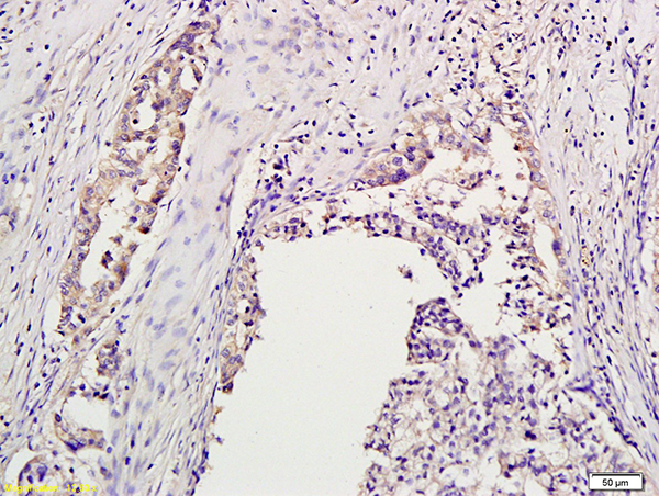
![IHC-P analysis of human prostate tissue using GTX18095 Aurora B antibody [AURKB/1593].](https://www.genetex.com/upload/website/prouct_img/normal/GTX18095/GTX18095_20200115_IHC-P_1055_w_23060620_221.webp)
Product group Antibodies
Aurora B antibody [AURKB/1593]GTX18095
ApplicationsImmunoHistoChemistry, ImmunoHistoChemistry Paraffin, Other Application
ReactivityHuman
TargetAURKB
- SizePrice
![Untreated (–) and treated (+) HeLa whole cell extracts (30 μg) were separated by 10% SDS-PAGE, and the membrane was blotted with Aurora B antibody [GT1146] (GTX00916) diluted at 1:500. The HRP-conjugated anti-rabbit IgG antibody (GTX213110-01) was used to detect the primary antibody.](https://www.genetex.com/upload/website/prouct_img/normal/GTX00916/GTX00916_40000000018_20200306_WB_treatment_Nocodazole_w_23053121_609.webp)
Product group Antibodies
Aurora B antibody [GT1146]GTX00916
ApplicationsImmunoFluorescence, Western Blot, ImmunoCytoChemistry
ReactivityHuman, Rat
TargetAURKB
- SizePrice