

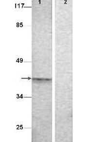
Western Blot shows detection of Aurora B protein at 39 kD (predicted band size).
All lanes: Aurora B (phospho T232) antibody diluted 1:500.
Lane 1: Extract from COS7 cells treated with Nocodazole (1 ug/ml, 16 hrs)
Lane 2: Extract from COS7 cells treated with Nocodazole (1 ug/ml, 16 hrs) and with the phosphopeptide immunogen
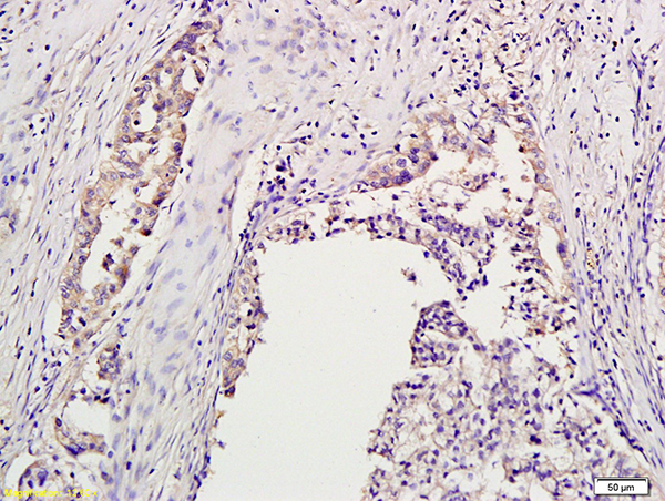
 stained with AURKB antibody (GTX85607) at 2.5 ug/ml followed by biotinylated goat anti-rabbit IgG secondary antibody LS-D1, alkaline phosphatase-streptavidin and chromogen.")
 using GTX85607 Aurora B (phospho Thr232) antibody. Antigen retrieval : not performed. Dilution : 10 μg/mL")
 using GTX85607 Aurora B (phospho Thr232) antibody. Antigen retrieval : not performed. Dilution : 10 μg/mL")
 antibody prior incubated with (Lane 2) or without (Lane 1) blocking peptide. Dilution : 1:500")
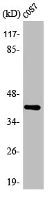
Western Blot shows detection of Aurora B protein at 39 kD (predicted band size).
All lanes: Aurora B (phospho T232) antibody diluted 1:500.
Lane 1: Extract from COS7 cells treated with Nocodazole (1 ug/ml, 16 hrs)
Lane 2: Extract from COS7 cells treated with Nocodazole (1 ug/ml, 16 hrs) and with the phosphopeptide immunogen
Aurora B (phospho Thr232) antibody
GTX85607
ApplicationsDot Blot, ImmunoFluorescence, Western Blot, ELISA, ImmunoCytoChemistry, ImmunoHistoChemistry, ImmunoHistoChemistry Paraffin, Other Application
Product group Antibodies
ReactivityHuman, Monkey
TargetAURKB
Overview
- SupplierGeneTex
- Product NameAurora B (phospho Thr232) antibody
- Delivery Days Customer9
- Application Supplier NoteWB: 1:250-1:2000. ELISA: 1:10000-1:30000. *Optimal dilutions/concentrations should be determined by the researcher.Not tested in other applications.
- ApplicationsDot Blot, ImmunoFluorescence, Western Blot, ELISA, ImmunoCytoChemistry, ImmunoHistoChemistry, ImmunoHistoChemistry Paraffin, Other Application
- CertificationResearch Use Only
- ClonalityPolyclonal
- Concentration0.87 mg/ml
- ConjugateUnconjugated
- Gene ID9212
- Target nameAURKB
- Target descriptionaurora kinase B
- Target synonymsAIK2, AIM-1, AIM1, ARK-2, ARK2, AurB, IPL1, PPP1R48, STK-1, STK12, STK5, aurkb-sv1, aurkb-sv2, aurora kinase B, aurora kinase B-Sv1, aurora kinase B-Sv2, aurora- and Ipl1-like midbody-associated protein 1, aurora- and Ipl1-like midbody-associated protein 1 homolog, aurora-1, aurora-B, aurora-related kinase 2, aurora/IPL1-related kinase 2, protein phosphatase 1, regulatory subunit 48, serine/threonine kinase 12, serine/threonine-protein kinase 12, serine/threonine-protein kinase 5, serine/threonine-protein kinase aurora-B
- HostRabbit
- IsotypeIgG
- Protein IDQ96GD4
- Protein NameAurora kinase B
- Scientific DescriptionChromosomal segregation during mitosis as well as meiosis is regulated by kinases and phosphatases. The Aurora kinases associate with microtubules during chromosome movement and segregation. Aurora kinase B localizes to microtubules near kinetochores, specifically to the specialized microtubules called K-fibers, and Aurora kinase A (MIM 603072) localizes to centrosomes (Lampson et al., 2004 [PubMed 14767480]).[supplied by OMIM]
- ReactivityHuman, Monkey
- Storage Instruction-20°C or -80°C,2°C to 8°C
- UNSPSC12352203
References
- Jagtap AD, Chang PT, Liu JR, et al. Novel acylureidoindolin-2-one derivatives as dual Aurora B/FLT3 inhibitors for the treatment of acute myeloid leukemia. Eur J Med Chem. 2014,85:268-88. doi: 10.1016/j.ejmech.2014.07.108Read this paper
- Wang HC, Jagtap AD, Chang PT, et al. Bioisosteric replacement of an acylureido moiety attached to an indolin-2-one scaffold with a malonamido or a 2/4-pyridinoylamido moiety produces a selectively potent Aurora-B inhibitor. Eur J Med Chem. 2014,84:312-34. doi: 10.1016/j.ejmech.2014.07.033Read this paper
Datasheet
Related products
Product group Antibodies
Aurkb Polyclonal AntibodyCAC07483
ApplicationsImmunoFluorescence, ELISA, ImmunoHistoChemistry
TargetAURKB
- SizePrice
Product group Antibodies
References
Aurora B Polyclonal AntibodyBS-2445R
ApplicationsFlow Cytometry, Western Blot, ELISA, ImmunoHistoChemistry, ImmunoHistoChemistry Paraffin
ReactivityBovine, Equine, Human, Mouse, Porcine, Rabbit, Rat
TargetAURKB
- SizePrice
Product group Antibodies
AURKB AntibodyCSB-PA000926
ApplicationsImmunoFluorescence, Western Blot, ELISA, ImmunoHistoChemistry
ReactivityHuman, Monkey, Mouse, Rat
TargetAURKB
- SizePrice
![IHC-P analysis of human kidney tissue section using GTX02594 Aurora B antibody [rAURKB/1592].](https://www.genetex.com/upload/website/prouct_img/normal/GTX02594/GTX02594_20210319_IHC-P_1_w_23053122_580.webp)
Product group Antibodies
Aurora B antibody [rAURKB/1592]GTX02594
ApplicationsImmunoHistoChemistry, ImmunoHistoChemistry Paraffin
ReactivityHuman
TargetAURKB
- SizePrice
![IHC-P analysis of human colon tissue section using GTX02595 Aurora B antibody [AURKB/3121R].](https://www.genetex.com/upload/website/prouct_img/normal/GTX02595/GTX02595_20210319_IHC-P_w_23053122_818.webp)
Product group Antibodies
Aurora B antibody [AURKB/3121R]GTX02595
ApplicationsImmunoHistoChemistry, ImmunoHistoChemistry Paraffin
ReactivityHuman
TargetAURKB
- SizePrice

Product group Antibodies
Anti-AURKB AntibodyHPA037708
ApplicationsImmunoCytoChemistry
ReactivityHuman
TargetAURKB
- SizePrice
Product group Antibodies
Aurora B antibodyGTX80313
ApplicationsWestern Blot
ReactivityHuman
TargetAURKB
- SizePrice