

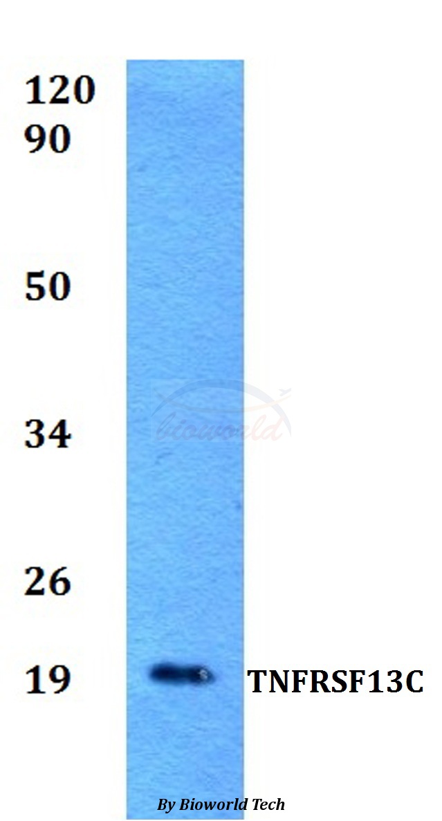
BAFF Receptor antibody
GTX31252
ApplicationsWestern Blot, ImmunoHistoChemistry, ImmunoHistoChemistry Paraffin
Product group Antibodies
ReactivityHuman, Mouse, Rat
TargetTNFRSF13C
Overview
- SupplierGeneTex
- Product NameBAFF Receptor antibody
- Delivery Days Customer9
- Application Supplier NoteWB: 2-5microg/ml. *Optimal dilutions/concentrations should be determined by the researcher.Not tested in other applications.
- ApplicationsWestern Blot, ImmunoHistoChemistry, ImmunoHistoChemistry Paraffin
- CertificationResearch Use Only
- ClonalityPolyclonal
- Concentration1 mg/ml
- ConjugateUnconjugated
- Gene ID115650
- Target nameTNFRSF13C
- Target descriptionTNF receptor superfamily member 13C
- Target synonymsBAFF-R, BAFFR, BROMIX, CD268, CVID4, prolixin, tumor necrosis factor receptor superfamily member 13C, B cell-activating factor receptor, BAFF receptor, BLyS receptor 3
- HostRabbit
- IsotypeIgG
- Protein IDQ96RJ3
- Protein NameTumor necrosis factor receptor superfamily member 13C
- Scientific DescriptionB cell-activating factor (BAFF) enhances B-cell survival in vitro and is a regulator of the peripheral B-cell population. Overexpression of Baff in mice results in mature B-cell hyperplasia and symptoms of systemic lupus erythematosus (SLE). Also, some SLE patients have increased levels of BAFF in serum. Therefore, it has been proposed that abnormally high levels of BAFF may contribute to the pathogenesis of autoimmune diseases by enhancing the survival of autoreactive B cells. The protein encoded by this gene is a receptor for BAFF and is a type III transmembrane protein containing a single extracellular cysteine-rich domain. It is thought that this receptor is the principal receptor required for BAFF-mediated mature B-cell survival. [provided by RefSeq, Jul 2008]
- ReactivityHuman, Mouse, Rat
- Storage Instruction-20°C or -80°C,2°C to 8°C
- UNSPSC12352203
Datasheet
Related products
Product group Antibodies

Anti-BR3 [V3-46s]Ab02649-10.0
ApplicationsELISA, Neutralisation/Blocking
ReactivityHuman, Mouse
TargetTNFRSF13C
- SizePrice
Product group Antibodies
Anti-TNFRSF13C Antibody144-63181
ApplicationsWestern Blot
ReactivityHuman, Mouse
TargetTNFRSF13C
- SizePrice
Product group Antibodies
ApplicationsWestern Blot, ELISA, ImmunoCytoChemistry, ImmunoHistoChemistry, ImmunoHistoChemistry Frozen, ImmunoHistoChemistry Paraffin
ReactivityPorcine
TargetTNFRSF13C
- SizePrice
Product group Antibodies
BAFFR Polyclonal AntibodyBS-2472R
ApplicationsFlow Cytometry, ImmunoFluorescence, ELISA, ImmunoCytoChemistry, ImmunoHistoChemistry, ImmunoHistoChemistry Frozen, ImmunoHistoChemistry Paraffin
ReactivityBovine, Canine, Equine, Human, Mouse, Rabbit, Rat
TargetTNFRSF13C
- SizePrice
Product group Antibodies
Anti-TNFRSF13C AntibodyA28703
ApplicationsWestern Blot
ReactivityHuman
- SizePrice
Product group Antibodies
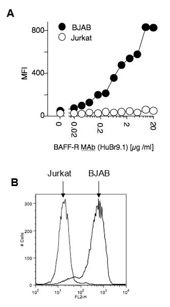
anti-BAFF-R (human), mAb (HuBR9.1)AG-20B-0016
ApplicationsFlow Cytometry
ReactivityHuman
TargetTNFRSF13C
- SizePrice

Product group Antibodies
Anti-TNFRSF13C AntibodyHPA003246
ApplicationsWestern Blot, ImmunoHistoChemistry
ReactivityHuman
TargetTNFRSF13C
- SizePrice
Product group Antibodies
BAFF Receptor antibodyGTX25965
ApplicationsWestern Blot, ImmunoHistoChemistry
ReactivityHuman, Mouse, Rat
TargetTNFRSF13C
- SizePrice