

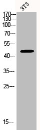
Whole cell extract (30 μg) was separated by 10% SDS-PAGE, and the membrane was blotted with BCKDK antibody [N2C3] (GTX104477) diluted at 1:1000. The HRP-conjugated anti-rabbit IgG antibody (GTX213110-01) was used to detect the primary antibody.
![BCKDK antibody [N2C3] detects BCKDK protein at mitochondria by immunofluorescent analysis. Sample: HeLa cells were fixed in 4% paraformaldehyde at RT for 15 min. Green: BCKDK stained by BCKDK antibody [N2C3] (GTX104477) diluted at 1:500. Red: alpha Tubulin, a cytoskeleton marker, stained by alpha Tubulin antibody [GT114] (GTX628802) diluted at 1:1000. Blue: Fluoroshield with DAPI (GTX30920).](https://www.genetex.com/upload/website/prouct_img/normal/GTX104477/GTX104477_44293_20221118_ICC_IF_22120719_839.webp "BCKDK antibody [N2C3] detects BCKDK protein at mitochondria by immunofluorescent analysis. Sample: HeLa cells were fixed in 4% paraformaldehyde at RT for 15 min. Green: BCKDK stained by BCKDK antibody [N2C3] (GTX104477) diluted at 1:500. Red: alpha Tubulin, a cytoskeleton marker, stained by alpha Tubulin antibody [GT114] (GTX628802) diluted at 1:1000. Blue: Fluoroshield with DAPI (GTX30920).")
![MCF-7 whole cell and membrane extracts (30 μg) were separated by 10% SDS-PAGE, and the membrane was blotted with BCKDK antibody [N2C3] (GTX104477) diluted at 1:1000. The HRP-conjugated anti-rabbit IgG antibody (GTX213110-01) was used to detect the primary antibody.](https://www.genetex.com/upload/website/prouct_img/normal/GTX104477/GTX104477_44510_20211203_WB_Fraction_23031402_372.webp "MCF-7 whole cell and membrane extracts (30 μg) were separated by 10% SDS-PAGE, and the membrane was blotted with BCKDK antibody [N2C3] (GTX104477) diluted at 1:1000. The HRP-conjugated anti-rabbit IgG antibody (GTX213110-01) was used to detect the primary antibody.")
 antibody at 1:100 dilution.
Antigen Retrieval: Trilogy? (EDTA based, pH 8.0) buffer, 15min")
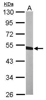
![Rat tissue extract (50 μg) was separated by 10% SDS-PAGE, and the membrane was blotted with BCKDK antibody [N2C3] (GTX104477) diluted at 1:10000. The HRP-conjugated anti-rabbit IgG antibody (GTX213110-01) was used to detect the primary antibody.](https://www.genetex.com/upload/website/prouct_img/normal/GTX104477/GTX104477_39785_20160804_WB_R_kidney_w_23060120_730.webp "Rat tissue extract (50 μg) was separated by 10% SDS-PAGE, and the membrane was blotted with BCKDK antibody [N2C3] (GTX104477) diluted at 1:10000. The HRP-conjugated anti-rabbit IgG antibody (GTX213110-01) was used to detect the primary antibody.")
![Wild-type (WT) and BCKDK knockout (KO) HeLa cell extracts (30 μg) were separated by 10% SDS-PAGE, and the membrane was blotted with BCKDK antibody [N2C3] (GTX104477) diluted at 1:1000. The HRP-conjugated anti-rabbit IgG antibody (GTX213110-01) was used to detect the primary antibody.](https://www.genetex.com/upload/website/prouct_img/normal/GTX104477/GTX104477_44293_20210507_WB_KO_watermark_w_23060120_669.webp "Wild-type (WT) and BCKDK knockout (KO) HeLa cell extracts (30 μg) were separated by 10% SDS-PAGE, and the membrane was blotted with BCKDK antibody [N2C3] (GTX104477) diluted at 1:1000. The HRP-conjugated anti-rabbit IgG antibody (GTX213110-01) was used to detect the primary antibody.")
![Rat tissue extract (50 μg) was separated by 10% SDS-PAGE, and the membrane was blotted with BCKDK antibody [N2C3] (GTX104477) diluted at 1:10000. The HRP-conjugated anti-rabbit IgG antibody (GTX213110-01) was used to detect the primary antibody.](https://www.genetex.com/upload/website/prouct_img/normal/GTX104477/GTX104477_39785_20160804_WB_R_brain_w_23060120_200.webp "Rat tissue extract (50 μg) was separated by 10% SDS-PAGE, and the membrane was blotted with BCKDK antibody [N2C3] (GTX104477) diluted at 1:10000. The HRP-conjugated anti-rabbit IgG antibody (GTX213110-01) was used to detect the primary antibody.")
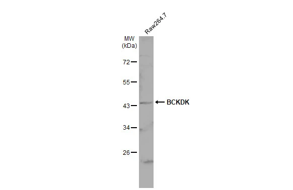
![Various whole cell extracts (30 μg) were separated by 10% SDS-PAGE, and the membrane was blotted with BCKDK antibody [N2C3] (GTX104477) diluted at 1:1000. The HRP-conjugated anti-rabbit IgG antibody (GTX213110-01) was used to detect the primary antibody.](https://www.genetex.com/upload/website/prouct_img/normal/GTX104477/GTX104477_44510_20231208_WB_23121122_140.webp "Various whole cell extracts (30 μg) were separated by 10% SDS-PAGE, and the membrane was blotted with BCKDK antibody [N2C3] (GTX104477) diluted at 1:1000. The HRP-conjugated anti-rabbit IgG antibody (GTX213110-01) was used to detect the primary antibody.")
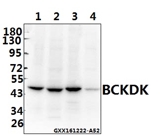
![Various tissue extracts (50 μg) were separated by 10% SDS-PAGE, and the membrane was blotted with BCKDK antibody [N2C3] (GTX104477) diluted at 1:10000. The HRP-conjugated anti-rabbit IgG antibody (GTX213110-01) was used to detect the primary antibody, and the signal was developed with Trident ECL plus-Enhanced.](https://www.genetex.com/upload/website/prouct_img/normal/GTX104477/GTX104477_44510_20231215_WB_M_R_23121922_189.webp "Various tissue extracts (50 μg) were separated by 10% SDS-PAGE, and the membrane was blotted with BCKDK antibody [N2C3] (GTX104477) diluted at 1:10000. The HRP-conjugated anti-rabbit IgG antibody (GTX213110-01) was used to detect the primary antibody, and the signal was developed with Trident ECL plus-Enhanced.")
Whole cell extract (30 μg) was separated by 10% SDS-PAGE, and the membrane was blotted with BCKDK antibody [N2C3] (GTX104477) diluted at 1:1000. The HRP-conjugated anti-rabbit IgG antibody (GTX213110-01) was used to detect the primary antibody.
BCKDK antibody [N2C3]
GTX104477
ApplicationsImmunoFluorescence, Western Blot, ImmunoCytoChemistry, ImmunoHistoChemistry, ImmunoHistoChemistry Paraffin
Product group Antibodies
ReactivityHuman, Mouse, Rat
TargetBCKDK
Overview
- SupplierGeneTex
- Product NameBCKDK antibody [N2C3]
- Delivery Days Customer9
- Application Supplier NoteWB: 1:500-1:20000. IHC-P: 1:100-1:1000. *Optimal dilutions/concentrations should be determined by the researcher.Not tested in other applications.
- ApplicationsImmunoFluorescence, Western Blot, ImmunoCytoChemistry, ImmunoHistoChemistry, ImmunoHistoChemistry Paraffin
- CertificationResearch Use Only
- ClonalityPolyclonal
- Concentration0.35 mg/ml
- ConjugateUnconjugated
- Gene ID10295
- Target nameBCKDK
- Target descriptionbranched chain keto acid dehydrogenase kinase
- Target synonymsBCKDKD, BDK, branched-chain alpha-ketoacid dehydrogenase kinase, BCKD-kinase, BCKDH kinase, BCKDHKIN, [3-methyl-2-oxobutanoate dehydrogenase [lipoamide]] kinase, mitochondrial, branched chain alpha-ketoacid dehydrogenase kinase, branched chain ketoacid dehydrogenase kinase
- HostRabbit
- IsotypeIgG
- Protein IDO14874
- Protein NameBranched-chain alpha-ketoacid dehydrogenase kinase
- Scientific DescriptionCatalyzes the phosphorylation and inactivation of the branched-chain alpha-ketoacid dehydrogenase complex, the key regulatory enzyme of the valine, leucine and isoleucine catabolic pathways. Key enzyme that regulate the activity state of the BCKD complex.
- ReactivityHuman, Mouse, Rat
- Storage Instruction-20°C or -80°C,2°C to 8°C
- UNSPSC41116161
Datasheet
Related products
Product group Antibodies
BCKDK AntibodyCSB-PA000981
ApplicationsImmunoFluorescence, Western Blot, ELISA, ImmunoHistoChemistry
ReactivityHuman, Mouse, Rat
TargetBCKDK
- SizePrice

Product group Antibodies
Anti-BCKDK Antibody Picoband(r)A07798-2-CARRIER-FREE
ApplicationsFlow Cytometry, ImmunoFluorescence, Western Blot, ELISA, ImmunoCytoChemistry, ImmunoHistoChemistry
ReactivityHuman, Mouse, Rat
TargetBCKDK
- SizePrice
Product group Antibodies
ApplicationsWestern Blot, ImmunoHistoChemistry
ReactivityHuman, Mouse, Rat
- SizePrice

Product group Antibodies
Anti-BCKDK AntibodyHPA017995
ApplicationsWestern Blot, ImmunoHistoChemistry
ReactivityHuman, Mouse, Rat
TargetBCKDK
- SizePrice
Product group Antibodies

BCKDK AntibodyLS-C409720
ApplicationsWestern Blot, ImmunoHistoChemistry
ReactivityHuman, Mouse, Rat
TargetBCKDK
- SizePrice
![Various tissue extracts (50 μg) were separated by 10% SDS-PAGE, and the membrane was blotted with BCKDK antibody [HL2710] (GTX639471) diluted at 1:1000. The HRP-conjugated anti-rabbit IgG antibody (GTX213110-01) was used to detect the primary antibody.](https://www.genetex.com/upload/website/prouct_img/normal/GTX639471/GTX639471_T-45250_20231215_WB_M_R_23121922_799.webp)
Product group Antibodies
BCKDK antibody [HL2710]GTX639471
ApplicationsWestern Blot, ImmunoHistoChemistry, ImmunoHistoChemistry Paraffin
ReactivityHuman, Mouse, Rat
TargetBCKDK
- SizePrice
![IHC-P analysis of human endometrium tissue using GTX84836 BCKDK antibody [11C9]. Antigen retrieval : Heat-induced epitope retrieval by 10mM citrate buffer, pH6.0, 100oC for 10min.](https://www.genetex.com/upload/website/prouct_img/normal/GTX84836/GTX84836_3156_IHC-P_w_23061420_542.webp)
Product group Antibodies
BCKDK antibody [11C9]GTX84836
ApplicationsImmunoFluorescence, Western Blot, ImmunoCytoChemistry, ImmunoHistoChemistry, ImmunoHistoChemistry Paraffin
ReactivityHuman
TargetBCKDK
- SizePrice
Product group Antibodies
BCKDK antibody [N1C2]GTX119413
ApplicationsWestern Blot, ImmunoHistoChemistry, ImmunoHistoChemistry Paraffin
ReactivityHuman, Mouse, Rat
TargetBCKDK
- SizePrice