

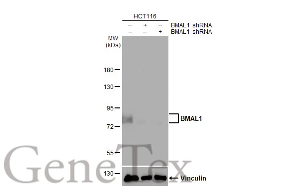
Non-transfected (–) and transfected (+) HCT-116 whole cell extract (30 μg) were separated by 7.5% SDS-PAGE, and the membrane was blotted with BMAL1 antibody [HL2456] (GTX638774) diluted at 1:1000. The HRP-conjugated anti-rabbit IgG antibody (GTX213110-01) was used to detect the primary antibody.
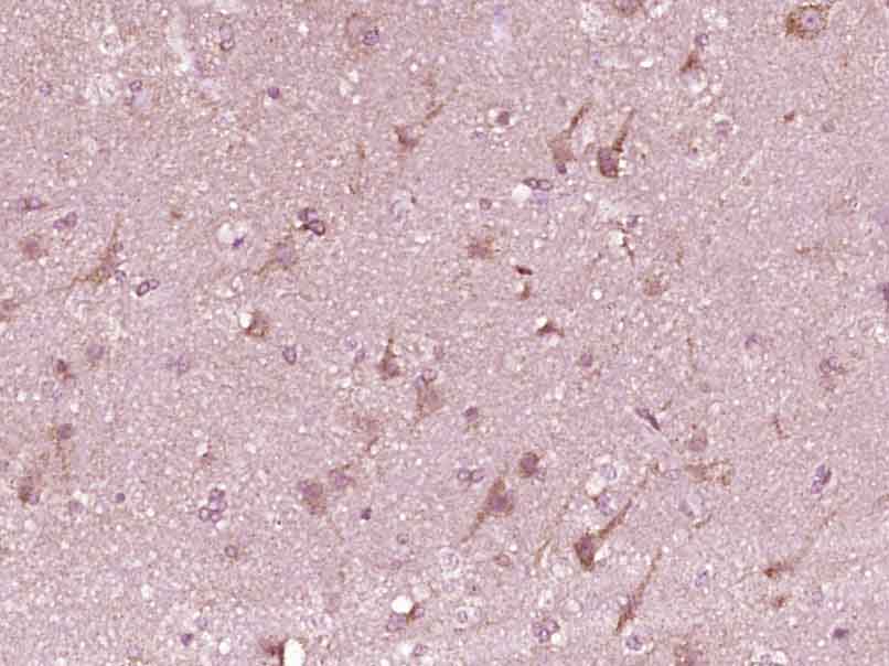
![BMAL1 antibody [HL2456] detects BMAL1 protein at cytoplasm and nucleus by immunohistochemical analysis. Sample: Paraffin-embedded rat intestine. BMAL1 stained by BMAL1 antibody [HL2456] (GTX638774) diluted at 1:100. Antigen Retrieval: Citrate buffer, pH 6.0, 15 min](https://www.genetex.com/upload/website/prouct_img/normal/GTX638774/GTX638774_T-45089_20230721_IHC-P_R_23073119_156.webp "BMAL1 antibody [HL2456] detects BMAL1 protein at cytoplasm and nucleus by immunohistochemical analysis. Sample: Paraffin-embedded rat intestine. BMAL1 stained by BMAL1 antibody [HL2456] (GTX638774) diluted at 1:100. Antigen Retrieval: Citrate buffer, pH 6.0, 15 min")
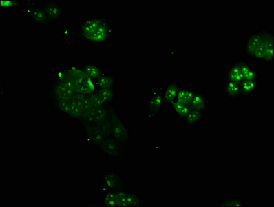
![BMAL1 antibody [HL2456] detects BMAL1 protein at cytoplasm and nucleus by immunofluorescent analysis. Sample: HCT-116 cells were fixed in 4% paraformaldehyde at RT for 15 min. Green: BMAL1 stained by BMAL1 antibody [HL2456] (GTX638774) diluted at 1:500. Red: alpha Tubulin, a cytoskeleton marker, stained by alpha Tubulin antibody [GT114] (GTX628802) diluted at 1:1000.](https://www.genetex.com/upload/website/prouct_img/normal/GTX638774/GTX638774_T-45089_20230804_ICC_IF_23080901_669.webp "BMAL1 antibody [HL2456] detects BMAL1 protein at cytoplasm and nucleus by immunofluorescent analysis. Sample: HCT-116 cells were fixed in 4% paraformaldehyde at RT for 15 min. Green: BMAL1 stained by BMAL1 antibody [HL2456] (GTX638774) diluted at 1:500. Red: alpha Tubulin, a cytoskeleton marker, stained by alpha Tubulin antibody [GT114] (GTX628802) diluted at 1:1000.")
![Various whole cell extracts (30 μg) were separated by 7.5% SDS-PAGE, and the membrane was blotted with BMAL1 antibody [HL2456] (GTX638774) diluted at 1:1000. The HRP-conjugated anti-rabbit IgG antibody (GTX213110-01) was used to detect the primary antibody, and the signal was developed with Trident ECL plus-Enhanced. Corresponding RNA expression data for the same cell lines are based on Human Protein Atlas program.](https://www.genetex.com/upload/website/prouct_img/normal/GTX638774/GTX638774_45159_20230908_WB_TPM_watermark_23091319_273.webp "Various whole cell extracts (30 μg) were separated by 7.5% SDS-PAGE, and the membrane was blotted with BMAL1 antibody [HL2456] (GTX638774) diluted at 1:1000. The HRP-conjugated anti-rabbit IgG antibody (GTX213110-01) was used to detect the primary antibody, and the signal was developed with Trident ECL plus-Enhanced. Corresponding RNA expression data for the same cell lines are based on Human Protein Atlas program.")
![Mouse tissue extract (50 μg) was separated by 7.5% SDS-PAGE, and the membrane was blotted with BMAL1 antibody [HL2456] (GTX638774) diluted at 1:1000. The HRP-conjugated anti-rabbit IgG antibody (GTX213110-01) was used to detect the primary antibody, and the signal was developed with Trident ECL plus-Enhanced.](https://www.genetex.com/upload/website/prouct_img/normal/GTX638774/GTX638774_45159_20231006_WB_M_brain_23102401_449.webp "Mouse tissue extract (50 μg) was separated by 7.5% SDS-PAGE, and the membrane was blotted with BMAL1 antibody [HL2456] (GTX638774) diluted at 1:1000. The HRP-conjugated anti-rabbit IgG antibody (GTX213110-01) was used to detect the primary antibody, and the signal was developed with Trident ECL plus-Enhanced.")
![Untreated and treated 293T whole cell extracts (30 μg) were separated by 7.5% SDS-PAGE, and the membrane was blotted with BMAL1 antibody [HL2456] (GTX638774) diluted at 1:1000. The HRP-conjugated anti-rabbit IgG antibody (GTX213110-01) was used to detect the primary antibody.](https://www.genetex.com/upload/website/prouct_img/normal/GTX638774/GTX638774_T-45089_20240223_WB_treatment_Serumshock_24022619_808.webp "Untreated and treated 293T whole cell extracts (30 μg) were separated by 7.5% SDS-PAGE, and the membrane was blotted with BMAL1 antibody [HL2456] (GTX638774) diluted at 1:1000. The HRP-conjugated anti-rabbit IgG antibody (GTX213110-01) was used to detect the primary antibody.")
![BMAL1 antibody [HL2456] detects BMAL1 protein by immunohistochemical analysis. Sample: Paraffin-embedded mouse suprachiasmatic nucleus. BMAL1 stained by BMAL1 antibody [HL2456] (GTX638774) diluted at 1:200. Antigen Retrieval: Tris-EDTA buffer, pH 9.0, 15 min](https://www.genetex.com/upload/website/prouct_img/normal/GTX638774/GTX638774_45159_20251114_IHC-P_M_25112800_249.webp "BMAL1 antibody [HL2456] detects BMAL1 protein by immunohistochemical analysis. Sample: Paraffin-embedded mouse suprachiasmatic nucleus. BMAL1 stained by BMAL1 antibody [HL2456] (GTX638774) diluted at 1:200. Antigen Retrieval: Tris-EDTA buffer, pH 9.0, 15 min")
Non-transfected (–) and transfected (+) HCT-116 whole cell extract (30 μg) were separated by 7.5% SDS-PAGE, and the membrane was blotted with BMAL1 antibody [HL2456] (GTX638774) diluted at 1:1000. The HRP-conjugated anti-rabbit IgG antibody (GTX213110-01) was used to detect the primary antibody.
BMAL1 antibody [HL2456]
GTX638774
ApplicationsImmunoFluorescence, Western Blot, ImmunoCytoChemistry, ImmunoHistoChemistry, ImmunoHistoChemistry Paraffin
Product group Antibodies
ReactivityHuman, Mouse, Rat
TargetBMAL1
Overview
- SupplierGeneTex
- Product NameBMAL1 antibody [HL2456]
- Delivery Days Customer9
- Application Supplier NoteWB: 1:500-1:3000. *Optimal dilutions/concentrations should be determined by the researcher.Not tested in other applications.
- ApplicationsImmunoFluorescence, Western Blot, ImmunoCytoChemistry, ImmunoHistoChemistry, ImmunoHistoChemistry Paraffin
- CertificationResearch Use Only
- ClonalityMonoclonal
- Clone IDHL2456
- Concentration1 mg/ml
- ConjugateUnconjugated
- Gene ID406
- Target nameBMAL1
- Target descriptionbasic helix-loop-helix ARNT like 1
- Target synonymsARNTL, ARNTL1, BMAL1c, JAP3, MOP3, PASD3, TIC, bHLHe5, basic helix-loop-helix ARNT-like protein 1, ARNT-like protein 1, brain and muscle, PAS domain containing 3, PAS domain-containing protein 3, aryl hydrocarbon receptor nuclear translocator like, aryl hydrocarbon receptor nuclear translocator-like protein 1, bHLH-PAS protein JAP3, basic helix-loop-helix family member e5, basic-helix-loop-helix-PAS orphan MOP3, basic-helix-loop-helix-PAS protein MOP3, brain and muscle ARNT-like 1, class E basic helix-loop-helix protein 5, member of PAS protein 3, member of PAS superfamily 3, mutant basic helix-loop-helix ARNT-like protein 1, testis tissue sperm-binding protein Li 50e
- HostRabbit
- IsotypeIgG
- Protein IDO00327
- Protein NameBasic helix-loop-helix ARNT-like protein 1
- Scientific DescriptionThe protein encoded by this gene is a basic helix-loop-helix protein that forms a heterodimer with CLOCK. This heterodimer binds E-box enhancer elements upstream of Period (PER1, PER2, PER3) and Cryptochrome (CRY1, CRY2) genes and activates transcription of these genes. PER and CRY proteins heterodimerize and repress their own transcription by interacting in a feedback loop with CLOCK/ARNTL complexes. Defects in this gene have been linked to infertility, problems with gluconeogenesis and lipogenesis, and altered sleep patterns. Several transcript variants encoding different isoforms have been found for this gene. [provided by RefSeq, Jul 2014]
- ReactivityHuman, Mouse, Rat
- Storage Instruction-20°C or -80°C,2°C to 8°C
- UNSPSC41116161
Datasheet
Related products
Product group Antibodies
ARNTL AntibodyCSB-PA002123LA01HU
ApplicationsImmunoFluorescence, ELISA
ReactivityHuman
TargetBMAL1
- SizePrice

Product group Antibodies
Anti-BMAL1/ARNTL Antibody Picoband(r)A00260-1-CARRIER-FREE
ApplicationsFlow Cytometry, Western Blot
ReactivityHuman, Mouse, Rat
TargetBMAL1
- SizePrice
Product group Antibodies
Anti-BMAL1 AntibodyA97688
ApplicationsWestern Blot, ELISA
ReactivityHuman, Mouse, Rat
- SizePrice
Product group Antibodies

Goat anti-BMAL1 / ARNTLEB09178
ApplicationsWestern Blot, ELISA
ReactivityBovine, Canine, Human, Mouse, Porcine, Rat
TargetBMAL1
- SizePrice

Product group Antibodies
Anti-ARNTL AntibodyHPA050938
ApplicationsImmunoCytoChemistry
ReactivityHuman
TargetBMAL1
- SizePrice
Product group Antibodies

ARNTL / BMAL1 AntibodyLS-C401453
ApplicationsWestern Blot, ELISA
ReactivityHuman, Mouse, Rat
TargetBMAL1
- SizePrice
Product group Antibodies
BMAL1 Polyclonal Antibodybs-3750R
ApplicationsFlow Cytometry, ImmunoFluorescence, Western Blot, ELISA, ImmunoCytoChemistry, ImmunoHistoChemistry, ImmunoHistoChemistry Frozen, ImmunoHistoChemistry Paraffin
ReactivityBovine, Canine, Equine, Human, Mouse, Porcine, Rat, Sheep
TargetBMAL1
- SizePrice
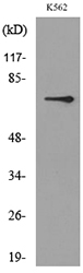
![WB analysis of BMAL1(AA: 1-310)-hIgGFc transfected HEK293 cell lysate using GTX83281 BMAL1 antibody [1C5].](https://www.genetex.com/upload/website/prouct_img/normal/GTX83281/GTX83281_20170912_WB_w_23061322_615.webp)
Product group Antibodies
BMAL1 antibody [1C5]GTX83281
ApplicationsWestern Blot, ELISA, ImmunoHistoChemistry, ImmunoHistoChemistry Paraffin
ReactivityHuman
TargetBMAL1
- SizePrice