

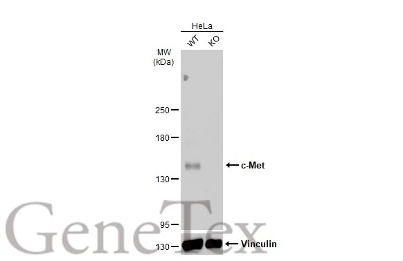
Wild-type (WT) and c-Met knockout (KO) HeLa cell extracts (30 μg) were separated by 5% SDS-PAGE, and the membrane was blotted with c-Met antibody [C3], C-term (GTX100637) diluted at 1:500. The HRP-conjugated anti-rabbit IgG antibody (GTX213110-01) was used to detect the primary antibody.
![Immunoprecipitation of c-Met protein from HeLa whole cell extracts using 5 μg of c-Met antibody [C3], C-term (GTX100637). Western blot analysis was performed using c-Met antibody [C3], C-term (GTX100637) diluted at 1:500. EasyBlot anti-Rabbit IgG (GTX221666-01) was used as a secondary reagent.](https://www.genetex.com/upload/website/prouct_img/normal/GTX100637/GTX100637_39988_IP_w_23060100_769.webp "Immunoprecipitation of c-Met protein from HeLa whole cell extracts using 5 μg of c-Met antibody [C3], C-term (GTX100637). Western blot analysis was performed using c-Met antibody [C3], C-term (GTX100637) diluted at 1:500. EasyBlot anti-Rabbit IgG (GTX221666-01) was used as a secondary reagent.")
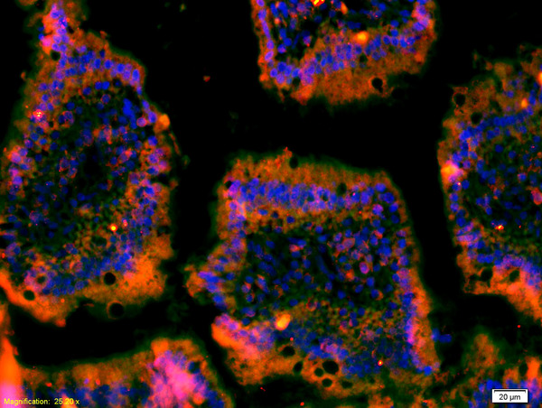
![c-Met antibody [c3], c-term detects c-Met protein at cytoplasm by immunofluorescent analysis. Sample: HeLa cells were fixed in 4% paraformaldehyde at RT for 15 min. Green: c-Met protein stained by c-Met antibody [c3], c-term (GTX100637) diluted at 1:1000. Red: alpha Tubulin, a cytoskeleton marker, stained by alpha Tubulin antibody [GT114] (GTX628802) diluted at 1:500. Blue: Hoechst 33342 staining.](https://www.genetex.com/upload/website/prouct_img/normal/GTX100637/GTX100637_39988_20150410_IFA_w_23060100_796.webp "c-Met antibody [c3], c-term detects c-Met protein at cytoplasm by immunofluorescent analysis. Sample: HeLa cells were fixed in 4% paraformaldehyde at RT for 15 min. Green: c-Met protein stained by c-Met antibody [c3], c-term (GTX100637) diluted at 1:1000. Red: alpha Tubulin, a cytoskeleton marker, stained by alpha Tubulin antibody [GT114] (GTX628802) diluted at 1:500. Blue: Hoechst 33342 staining.")
![Non-transfected (–) and transfected (+) HeLa whole cell extracts (30 μg) were separated by 5% SDS-PAGE, and the membrane was blotted with c-Met antibody [C3], C-term (GTX100637) diluted at 1:500. The HRP-conjugated anti-rabbit IgG antibody (GTX213110-01) was used to detect the primary antibody.](https://www.genetex.com/upload/website/prouct_img/normal/GTX100637/GTX100637_39988_20160901_WB_shRNA_watermark_w_23060100_148.webp "Non-transfected (–) and transfected (+) HeLa whole cell extracts (30 μg) were separated by 5% SDS-PAGE, and the membrane was blotted with c-Met antibody [C3], C-term (GTX100637) diluted at 1:500. The HRP-conjugated anti-rabbit IgG antibody (GTX213110-01) was used to detect the primary antibody.")
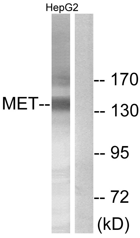
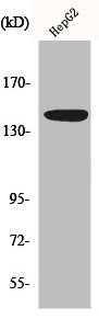
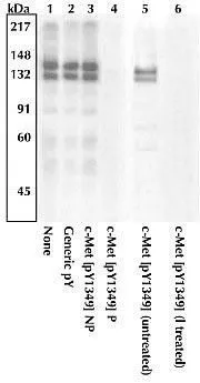
![Various whole cell extracts (30 μg) were separated by 5% SDS-PAGE, and the membrane was blotted with c-Met antibody [C3], C-term (GTX100637) diluted at 1:500. The HRP-conjugated anti-rabbit IgG antibody (GTX213110-01) was used to detect the primary antibody, and the signal was developed with Trident ECL plus-Enhanced.](https://www.genetex.com/upload/website/prouct_img/normal/GTX100637/GTX100637_43901_20200424_WB_w_23060100_713.webp "Various whole cell extracts (30 μg) were separated by 5% SDS-PAGE, and the membrane was blotted with c-Met antibody [C3], C-term (GTX100637) diluted at 1:500. The HRP-conjugated anti-rabbit IgG antibody (GTX213110-01) was used to detect the primary antibody, and the signal was developed with Trident ECL plus-Enhanced.")

Wild-type (WT) and c-Met knockout (KO) HeLa cell extracts (30 μg) were separated by 5% SDS-PAGE, and the membrane was blotted with c-Met antibody [C3], C-term (GTX100637) diluted at 1:500. The HRP-conjugated anti-rabbit IgG antibody (GTX213110-01) was used to detect the primary antibody.
c-Met antibody [C3], C-term
GTX100637
ApplicationsImmunoFluorescence, ImmunoPrecipitation, Western Blot, ImmunoCytoChemistry
Product group Antibodies
ReactivityHuman
TargetMET
Overview
- SupplierGeneTex
- Product Namec-Met antibody [C3], C-term
- Delivery Days Customer9
- Application Supplier NoteWB: 1:500-1:3000. ICC/IF: 1:100-1:1000. IP: 1:100-1:500. *Optimal dilutions/concentrations should be determined by the researcher.Not tested in other applications.
- ApplicationsImmunoFluorescence, ImmunoPrecipitation, Western Blot, ImmunoCytoChemistry
- CertificationResearch Use Only
- ClonalityPolyclonal
- Concentration1.95 mg/ml
- ConjugateUnconjugated
- Gene ID4233
- Target nameMET
- Target descriptionMET proto-oncogene, receptor tyrosine kinase
- Target synonymsAUTS9, DA11, DFNB97, HGFR, RCCP2, c-Met, hepatocyte growth factor receptor, HGF receptor, HGF/SF receptor, SF receptor, proto-oncogene c-Met, scatter factor receptor, tyrosine-protein kinase Met
- HostRabbit
- IsotypeIgG
- Protein IDP08581
- Protein NameHepatocyte growth factor receptor
- Scientific DescriptionThe proto-oncogene MET product is the hepatocyte growth factor receptor and encodes tyrosine-kinase activity. The primary single chain precursor protein is post-translationally cleaved to produce the alpha and beta subunits, which are disulfide linked to form the mature receptor. Various mutations in the MET gene are associated with papillary renal carcinoma. Two transcript variants encoding different isoforms have been found for this gene. [provided by RefSeq]
- ReactivityHuman
- Storage Instruction-20°C or -80°C,2°C to 8°C
- UNSPSC41116161
Datasheet
Related products
Product group Antibodies

c-Met (Phospho-Tyr1003) AntibodyABX012477
ApplicationsImmunoFluorescence, Western Blot, ELISA, ImmunoCytoChemistry, ImmunoHistoChemistry
- SizePrice
Product group Antibodies
Anti-Met AntibodyA101612
ApplicationsWestern Blot, ELISA
ReactivityHuman
- SizePrice
Product group Antibodies

Anti-MET [7A2]AB03829-23.0
ApplicationsELISA, Neutralisation/Blocking, Other Application
ReactivityHuman
TargetMET
- SizePrice
Product group Antibodies
References
MET Polyclonal AntibodyBS-0668R
ApplicationsImmunoFluorescence, Western Blot, ELISA, ImmunoHistoChemistry, ImmunoHistoChemistry Frozen, ImmunoHistoChemistry Paraffin
ReactivityHuman, Mouse, Rat
TargetMET
- SizePrice
Product group Antibodies
MET AntibodyCSB-PA003231
ApplicationsWestern Blot, ELISA
ReactivityHuman
TargetMET
- SizePrice
Product group Antibodies
C-Met (MET) Polyclonal AntibodyCAU24571
ApplicationsWestern Blot, ImmunoHistoChemistry
ReactivityMouse
TargetMET
- SizePrice
![WB analysis of various samples using GTX14700 c-Met antibody [3i20(25H2)]. Lane 1 : mIMCD3 Lane 2 : Vero Lane 3 : 293 (HGF stimulated) Lane 4 : 293 (starved) Lane 5 : C6](https://www.genetex.com/upload/website/prouct_img/normal/GTX14700/GTX14700_20191203_WB_46_w_23060620_293.webp)
Product group Antibodies
c-Met antibody [3i20(25H2)]GTX14700
ApplicationsImmunoPrecipitation, Western Blot, ImmunoHistoChemistry
ReactivityHuman, Monkey, Mouse, Primate, Rat
TargetMET
- SizePrice
Product group Antibodies
c-Met (phospho Tyr1349) antibodyGTX25669
ApplicationsWestern Blot
ReactivityHuman
TargetMET
- SizePrice