

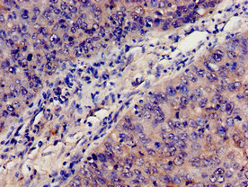
Immunohistochemistry analysis of human cervical cancer using CSB-PA10599A0Rb at dilution of 1:100

")
. Section was blocked with 10% normal goat serum 30min at RT. Then primary antibody (1% BSA) was incubated at 4°C overnight. The primary is detected by a biotinylated secondary antibody and visualized using an HRP conjugated SP system.")
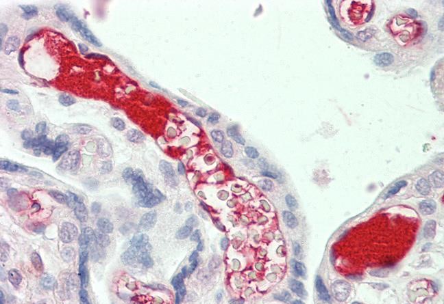
. Section was blocked with 10% normal goat serum 30min at RT. Then primary antibody (1% BSA) was incubated at 4°C overnight. The primary is detected by a biotinylated secondary antibody and visualized using an HRP conjugated SP system.")
Immunohistochemistry analysis of human cervical cancer using CSB-PA10599A0Rb at dilution of 1:100
C3 Antibody
CSB-PA10599A0RB
ApplicationsImmunoFluorescence, ELISA, ImmunoHistoChemistry
Product group Antibodies
ReactivityHuman
TargetC3
Overview
- SupplierCusabio
- Product NameC3 Antibody
- Delivery Days Customer20
- ApplicationsImmunoFluorescence, ELISA, ImmunoHistoChemistry
- CertificationResearch Use Only
- ClonalityPolyclonal
- ConjugateUnconjugated
- Gene ID718
- Target nameC3
- Target descriptioncomplement C3
- Target synonymsAHUS5, ARMD9, ASP, C3a, C3b, CPAMD1, HEL-S-62p, complement C3, C3 and PZP-like alpha-2-macroglobulin domain-containing protein 1, C3a anaphylatoxin, acylation-stimulating protein cleavage product, complement component 3, complement component C3a, complement component C3b, epididymis secretory sperm binding protein Li 62p, prepro-C3
- HostRabbit
- IsotypeIgG
- Protein IDP01024
- Protein NameComplement C3
- Scientific DescriptionC3 plays a central role in the activation of the complement system. Its processing by C3 convertase is the central reaction in both classical and alternative complement pathways. After activation C3b can bind covalently, via its reactive thioester, to cell surface carbohydrates or immune aggregates. Ref.5 Ref.11 Ref.14 Ref.15 Ref.16 Ref.22 Ref.25 Ref.30 Derived from proteolytic degradation of complement C3, C3a anaphylatoxin is a mediator of local inflammatory process. It induces the contraction of smooth muscle, increases vascular permeability and causes histamine release from mast cells and basophilic leukocytes. Ref.5 Ref.11 Ref.14 Ref.15 Ref.16 Ref.22 Ref.25 Ref.30 Acylation stimulating protein (ASP): adipogenic hormone that stimulates triglyceride (TG) synthesis and glucose transport in adipocytes, regulating fat storage and playing a role in postprandial TG clearance. Appears to stimulate TG synthesis via activation of the PLC, MAPK and AKT signaling pathways. Ligand for GPR77. Promotes the phosphorylation, ARRB2-mediated internalization and recycling of GPR77.
- ReactivityHuman
- Storage Instruction-20°C or -80°C
- UNSPSC41116161
Related products
Product group Antibodies
Anti-C3 AntibodyA286077
ApplicationsELISA, ImmunoHistoChemistry
ReactivityHuman
- SizePrice
Product group Antibodies

Anti-C3b-iC3b [7C12]Ab01554-1.1
ApplicationsFlow Cytometry, ImmunoFluorescence, Western Blot, ELISA
ReactivityHuman
TargetC3
- SizePrice

Product group Antibodies
Anti-C3 Antibody Picoband(r)A00168-3-CARRIER-FREE
ApplicationsFlow Cytometry, Western Blot, ELISA
ReactivityHuman, Mouse, Rat
TargetC3
- SizePrice
Product group Antibodies

ApplicationsELISA
ReactivityHuman
TargetC3
- SizePrice
Product group Antibodies
C3 Polyclonal AntibodyCAC09129
ApplicationsImmunoFluorescence, ELISA, ImmunoHistoChemistry
TargetC3
- SizePrice
Product group Antibodies
References
ApplicationsImmunoFluorescence, Western Blot, ELISA, ImmunoHistoChemistry, ImmunoHistoChemistry Frozen, ImmunoHistoChemistry Paraffin
ReactivityHuman
TargetC3
- SizePrice
![C3 antibody [C3], C-term detects C3 protein at cytoplasm by immunofluorescent analysis. Sample: HeLa cells were fixed in 4% paraformaldehyde at RT for 15 min. Green: C3 protein stained by C3 antibody [C3], C-term (GTX101316) diluted at 1:200. Blue: Hoechst 33342 staining.](https://www.genetex.com/upload/website/prouct_img/normal/GTX101316/GTX101316_42116_20150924_IFA_w_23060100_328.webp)
Product group Antibodies
C3 / C3b antibody [C3], C-termGTX101316
ApplicationsImmunoFluorescence, ImmunoPrecipitation, Western Blot, ImmunoCytoChemistry, ImmunoHistoChemistry, ImmunoHistoChemistry Paraffin, Other Application
ReactivityHuman, Mouse
TargetC3
- SizePrice

Product group Antibodies
Anti-C3 AntibodyHPA003563
ApplicationsImmunoHistoChemistry
ReactivityHuman
TargetC3
- SizePrice