

Anti-C3b-iC3b [7C12]
Ab01554-1.1
ApplicationsFlow Cytometry, ImmunoFluorescence, Western Blot, ELISA
Product group Antibodies
ReactivityHuman
TargetC3
Overview
- SupplierAbsolute Antibody
- Product NameAnti-C3b-iC3b [7C12]
- Delivery Days Customer7
- Application Supplier NoteTo prepare and characterize this antibody, an ELISA on iC3 protein was performed using the mouse version of this antibody (Tosic et al, 1989; pmid:2786912). An immunoblot was performed on control, early AD, and AD patient brain tissues using the mouse version of this antibody to determine the link between the complement system and Alzheimers disease (Wu et al, 2019; pmid:31433986). To study the deposition of the complement protein fragment C3b and its breakdown products on CD20-positive cells, flow cytometry was performed on Raji or ARH-77 cells using the mouse version of this antibody. Furthermore, a radioimmunoassay was performed on Raji or ARH-77 cells using this antibody. Finally, Raji or ARH-77 cells were stained for immunofluorescence using this antibody (Kennedy et al, 2003; pmid:12393727). To study how Rituximab activates complement and promotes covalent deposition of C3 fragments (C3b/iC3b) on cells, flow cytometry was performed on raji cells in presence of the mouse version of this antibody. Furthermore, raji cells were stained for immunofluorescence using this antibody (Beum et al, 2006; pmid:17067631). To study the alternative pathway (AP) of the complement system, flow cytometry was performed on AP-mediated C3b-opsonizated zymosan using the mouse version of this antibody. Furthermore, immunofluorescence was performed on AP-mediated C3b-opsonizated zymosan using this antibody (DiLillo et al, 2006; pmid:15961157).
- ApplicationsFlow Cytometry, ImmunoFluorescence, Western Blot, ELISA
- Applications SupplierELISA; WB; IF; FC
- CertificationResearch Use Only
- ClonalityMonoclonal
- Clone ID7C12
- Gene ID718
- Target nameC3
- Target descriptioncomplement C3
- Target synonymsAHUS5, ARMD9, ASP, C3a, C3b, CPAMD1, HEL-S-62p, complement C3, C3 and PZP-like alpha-2-macroglobulin domain-containing protein 1, C3a anaphylatoxin, acylation-stimulating protein cleavage product, complement component 3, complement component C3a, complement component C3b, epididymis secretory sperm binding protein Li 62p, prepro-C3
- HostMouse
- IsotypeIgG1
- Protein IDP01024
- Protein NameComplement C3
- ReactivityHuman
- Reactivity SupplierHuman
- Reactivity Supplier NoteThe mouse version of this antibody was raised by immunizing a mouse with human C3b and iC3b bound to a carbohydrate matrix via the alternative pathway of complement activation.
- Storage Instruction-20°C,2°C to 8°C
- UNSPSC41116161
Related products
Product group Antibodies
Anti-C3 AntibodyA286077
ApplicationsELISA, ImmunoHistoChemistry
ReactivityHuman
- SizePrice

Product group Antibodies
Anti-C3 Antibody Picoband(r)A00168-3-CARRIER-FREE
ApplicationsFlow Cytometry, Western Blot, ELISA
ReactivityHuman, Mouse, Rat
TargetC3
- SizePrice
Product group Antibodies
C3 AntibodyCSB-PA10599A0RB
ApplicationsImmunoFluorescence, ELISA, ImmunoHistoChemistry
ReactivityHuman
TargetC3
- SizePrice
Product group Antibodies

ApplicationsELISA
ReactivityHuman
TargetC3
- SizePrice
Product group Antibodies
C3 Polyclonal AntibodyCAC09129
ApplicationsImmunoFluorescence, ELISA, ImmunoHistoChemistry
TargetC3
- SizePrice
Product group Antibodies
References
ApplicationsImmunoFluorescence, Western Blot, ELISA, ImmunoHistoChemistry, ImmunoHistoChemistry Frozen, ImmunoHistoChemistry Paraffin
ReactivityHuman
TargetC3
- SizePrice
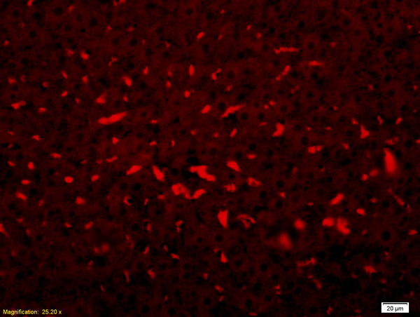
![C3 antibody [C3], C-term detects C3 protein at cytoplasm by immunofluorescent analysis. Sample: HeLa cells were fixed in 4% paraformaldehyde at RT for 15 min. Green: C3 protein stained by C3 antibody [C3], C-term (GTX101316) diluted at 1:200. Blue: Hoechst 33342 staining.](https://www.genetex.com/upload/website/prouct_img/normal/GTX101316/GTX101316_42116_20150924_IFA_w_23060100_328.webp)
Product group Antibodies
C3 / C3b antibody [C3], C-termGTX101316
ApplicationsImmunoFluorescence, ImmunoPrecipitation, Western Blot, ImmunoCytoChemistry, ImmunoHistoChemistry, ImmunoHistoChemistry Paraffin, Other Application
ReactivityHuman, Mouse
TargetC3
- SizePrice

Product group Antibodies
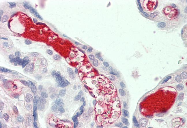
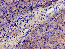
Anti-C3 AntibodyHPA003563
ApplicationsImmunoHistoChemistry
ReactivityHuman
TargetC3
- SizePrice