

Various tissue extracts (50 μg) were separated by 7.5% SDS-PAGE, and the membrane was blotted with Calnexin antibody (GTX112886) diluted at 1:1000. The HRP-conjugated anti-rabbit IgG antibody (GTX213110-01) was used to detect the primary antibody.
 and transfected (+) 293T whole cell extracts (30 μg) were separated by 7.5% SDS-PAGE, and the membrane was blotted with Calnexin antibody (GTX112886) diluted at 1:5000. The HRP-conjugated anti-rabbit IgG antibody (GTX213110-01) was used to detect the primary antibody.")
 A: mouse liver 7.5% SDS PAGE GTX112886 diluted at 1:1000")
 antibody at 1:500 dilution.
Antigen Retrieval: Trilogy? (EDTA based, pH 8.0) buffer, 15min")
 diluted at 1:500.
Antigen Retrieval: Citrate buffer, pH 6.0, 15 min")
 A: 293T B: A431 C: HeLa D: HepG2 7.5% SDS PAGE GTX112886 diluted at 1:10000")
 and transfected (+) 293T whole cell extracts (15 μg) were separated by 7.5% SDS-PAGE, and the membrane was blotted with Calnexin antibody (GTX112886) diluted at 1:10000.")
 diluted at 1:200. Blue: Hoechst 33342 staining.")
 were separated by 7.5% SDS-PAGE, and the membrane was blotted with Calnexin antibody (GTX112886) diluted at 1:1000.")
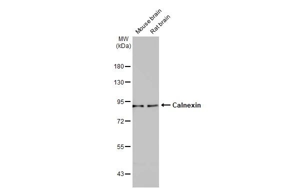
Various tissue extracts (50 μg) were separated by 7.5% SDS-PAGE, and the membrane was blotted with Calnexin antibody (GTX112886) diluted at 1:1000. The HRP-conjugated anti-rabbit IgG antibody (GTX213110-01) was used to detect the primary antibody.
Calnexin antibody
GTX112886
ApplicationsImmunoFluorescence, Western Blot, ImmunoCytoChemistry, ImmunoHistoChemistry, ImmunoHistoChemistry Paraffin
Product group Antibodies
ReactivityHuman, Mouse, Rat
TargetCANX
Overview
- SupplierGeneTex
- Product NameCalnexin antibody
- Delivery Days Customer9
- Application Supplier NoteWB: 1:500-1:10000. ICC/IF: 1:100-1:1000. IHC-P: 1:100-1:1000. *Optimal dilutions/concentrations should be determined by the researcher.Not tested in other applications.
- ApplicationsImmunoFluorescence, Western Blot, ImmunoCytoChemistry, ImmunoHistoChemistry, ImmunoHistoChemistry Paraffin
- CertificationResearch Use Only
- ClonalityPolyclonal
- Concentration0.53 mg/ml
- ConjugateUnconjugated
- Gene ID821
- Target nameCANX
- Target descriptioncalnexin
- Target synonymsCNX, IP90, P90, calnexin, epididymis secretory sperm binding protein, major histocompatibility complex class I antigen-binding protein p88
- HostRabbit
- IsotypeIgG
- Protein IDP27824
- Protein NameCalnexin
- Scientific DescriptionThis gene encodes a member of the calnexin family of molecular chaperones. The encoded protein is a calcium-binding, endoplasmic reticulum (ER)-associated protein that interacts transiently with newly synthesized N-linked glycoproteins, facilitating protein folding and assembly. It may also play a central role in the quality control of protein folding by retaining incorrectly folded protein subunits within the ER for degradation. Alternatively spliced transcript variants encoding the same protein have been described. [provided by RefSeq]
- ReactivityHuman, Mouse, Rat
- Storage Instruction-20°C or -80°C,2°C to 8°C
- UNSPSC12352203
References
- Pendiuk Goncalves J, Cruz Villarreal J, Walker SA, et al. High-throughput analysis of glycan sorting into extracellular vesicles. Biochim Biophys Acta Mol Cell Res. 2024,1871(2):119641. doi: 10.1016/j.bbamcr.2023.119641Read this paper
- Guo P, Busatto S, Huang J, et al. A facile magnetic extrusion method for preparing endosome-derived vesicles for cancer drug delivery. Adv Funct Mater. 2021,31(44):pii: 2008326. doi: 10.1002/adfm.202008326.Read this paper
- Mun SJ, Kang SA, Park HK, et al. Intranasally Administered Extracellular Vesicles from Adipose Stem Cells Have Immunomodulatory Effects in a Mouse Model of Asthma. Stem Cells Int. 2021,2021:6686625. doi: 10.1155/2021/6686625Read this paper
- Busatto S, Iannotta D, Walker SA, et al. A Simple and Quick Method for Loading Proteins in Extracellular Vesicles. Pharmaceuticals (Basel). 2021,14(4). doi: 10.3390/ph14040356Read this paper
- Miyauchi Y, Tanaka Y, Nagata K, et al. UDP-Glucuronosyltransferase (UGT)-mediated attenuations of cytochrome P450 3A4 activity: UGT isoform-dependent mechanism of suppression. Br J Pharmacol. 2020,177(5):1077-1089. doi: 10.1111/bph.14900Read this paper
- Huang HJ, Lin CC, Chou HC, et al. Proteomic analysis of rhein-induced cyt: ER stress mediates cell death in breast cancer cells. Mol Biosyst. 2014,10(12):3086-100. doi: 10.1039/c4mb00451eRead this paper
Datasheet
Related products
Product group Antibodies
Anti-CANX Antibody144-63846
ApplicationsImmunoFluorescence, Western Blot, ImmunoHistoChemistry
ReactivityHuman, Mouse, Rat
TargetCANX
- SizePrice

Product group Antibodies
Anti-Calnexin/CANX Antibody Picoband(r)A03372-2-CARRIER-FREE
ApplicationsFlow Cytometry, ImmunoFluorescence, Western Blot, ELISA, ImmunoCytoChemistry, ImmunoHistoChemistry
ReactivityHuman, Mouse
TargetCANX
- SizePrice
![Various whole cell extracts (30 μg) were separated by 7.5% SDS-PAGE, and the membrane was blotted with Calnexin antibody [N3C2], Internal (GTX101676) diluted at 1:5000. The HRP-conjugated anti-rabbit IgG antibody (GTX213110-01) was used to detect the primary antibody.](https://www.genetex.com/upload/website/prouct_img/normal/GTX101676/GTX101676_43643_20190719_WB_w_23060100_608.webp)
Product group Antibodies
References
Calnexin antibody [N3C2], InternalGTX101676
ApplicationsImmunoFluorescence, Western Blot, ImmunoCytoChemistry, ImmunoHistoChemistry, ImmunoHistoChemistry Paraffin
ReactivityHuman, Mouse, Rat
TargetCANX
- SizePrice
![Calnexin antibody [C3], C-term detects Calnexin protein at cell membrane and cytoplasm by immunohistochemical analysis. Sample: Paraffin-embedded rat brain. Calnexin stained by Calnexin antibody [C3], C-term (GTX109669) diluted at 1:165. Antigen Retrieval: Citrate buffer, pH 6.0, 15 min](https://www.genetex.com/upload/website/prouct_img/normal/GTX109669/GTX109669_44699_20220701_IHC-P_R_22071401_447.webp)
Product group Antibodies
References
Calnexin antibody [C3], C-termGTX109669
ApplicationsImmunoFluorescence, ImmunoPrecipitation, Western Blot, ImmunoCytoChemistry, ImmunoHistoChemistry, ImmunoHistoChemistry Paraffin
ReactivityHuman, Mouse, Rat, Sheep
TargetCANX
- SizePrice
Product group Antibodies
Calnexin antibody [AT18B9]GTX57717
ApplicationsFlow Cytometry, ImmunoFluorescence, Western Blot, ImmunoCytoChemistry
ReactivityHuman
TargetCANX
- SizePrice
![Calnexin antibody [GT1563] detects Calnexin protein at cytoplasm by immunofluorescent analysis. Sample: Neuro2A cells were fixed in 4% paraformaldehyde at RT for 15 min. Green: Calnexin stained by Calnexin antibody [GT1563] (GTX629976) diluted at 1:500. Blue: Fluoroshield with DAPI (GTX30920).](https://www.genetex.com/upload/website/prouct_img/normal/GTX629976/GTX629976_41484_20230512_ICC_IF_M_23060622_501.webp)
Product group Antibodies
References
Calnexin antibody [GT1563]GTX629976
ApplicationsDot Blot, ImmunoFluorescence, ImmunoPrecipitation, Western Blot, ImmunoCytoChemistry
ReactivityHuman, Mouse, Rat
TargetCANX
- SizePrice
![Calnexin antibody [HL1598] detects Calnexin protein at cytoplasm by immunohistochemical analysis. Sample: Paraffin-embedded mouse intestine. Calnexin stained by Calnexin antibody [HL1598] (GTX637077) diluted at 1:100. Antigen Retrieval: Citrate buffer, pH 6.0, 15 min](https://www.genetex.com/upload/website/prouct_img/normal/GTX637077/GTX637077_T-44725_20220722_IHC-P_M_22080119_403.webp)
Product group Antibodies
Calnexin antibody [HL1598]GTX637077
ApplicationsWestern Blot, ImmunoHistoChemistry, ImmunoHistoChemistry Paraffin
ReactivityCanine, Feline, Human, Mouse, Rat
TargetCANX
- SizePrice
Product group Antibodies
CANX Polyclonal AntibodyCAC14640
ApplicationsImmunoFluorescence, Western Blot, ELISA, ImmunoHistoChemistry
TargetCANX
- SizePrice
Product group Antibodies
References
Calnexin Polyclonal AntibodyBS-1693R
ApplicationsFlow Cytometry, ImmunoFluorescence, Western Blot, ELISA, ImmunoCytoChemistry, ImmunoHistoChemistry, ImmunoHistoChemistry Frozen, ImmunoHistoChemistry Paraffin
ReactivityBovine, Chicken, Equine, Human, Mouse, Rabbit, Rat
TargetCANX
- SizePrice
Product group Antibodies
CANX AntibodyCSB-PA001175
ApplicationsImmunoFluorescence, Western Blot, ELISA, ImmunoHistoChemistry
ReactivityHuman, Mouse, Rat
TargetCANX
- SizePrice