

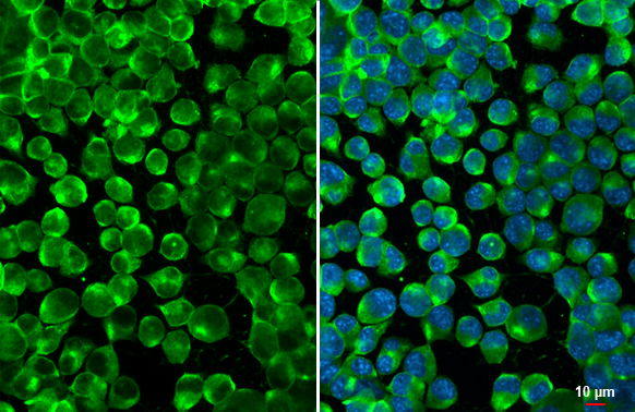
Calnexin antibody [GT1563] detects Calnexin protein at cytoplasm by immunofluorescent analysis. Sample: Neuro2A cells were fixed in 4% paraformaldehyde at RT for 15 min. Green: Calnexin stained by Calnexin antibody [GT1563] (GTX629976) diluted at 1:500. Blue: Fluoroshield with DAPI (GTX30920).
![Calnexin antibody [GT1563] detects Calnexin protein by Western blot analysis. A. 30 μg PC-12 whole cell lysate/extract B. 30 μg Rat-2 whole cell lysate/extract 7.5 % SDS-PAGE Calnexin antibody [GT1563] (GTX629976) dilution: 1:500](https://www.genetex.com/upload/website/prouct_img/normal/GTX629976/GTX629976_41484_WB_R_w_23061202_223.webp "Calnexin antibody [GT1563] detects Calnexin protein by Western blot analysis. A. 30 μg PC-12 whole cell lysate/extract B. 30 μg Rat-2 whole cell lysate/extract 7.5 % SDS-PAGE Calnexin antibody [GT1563] (GTX629976) dilution: 1:500")
![Non-transfected (–) and transfected (+) 293T whole cell extracts (15 μg) were separated by 7.5% SDS-PAGE, and the membrane was blotted with Calnexin antibody [GT1563] (GTX629976) diluted at 1:6000.](https://www.genetex.com/upload/website/prouct_img/normal/GTX629976/GTX629976_41484_20160707_WB_shRNA_watermark_w_23061202_617.webp "Non-transfected (–) and transfected (+) 293T whole cell extracts (15 μg) were separated by 7.5% SDS-PAGE, and the membrane was blotted with Calnexin antibody [GT1563] (GTX629976) diluted at 1:6000.")
![Calnexin antibody [GT1563] detects Calnexin protein by Western blot analysis. A. 30 μg 293T whole cell lysate/extract B. 30 μg A431 whole cell lysate/extract C. 30 μg HeLa whole cell lysate/extract D. 30 μg HepG2 whole cell lysate/extract 7.5 % SDS-PAGE Calnexin antibody [GT1563] (GTX629976) dilution: 1:1000](https://www.genetex.com/upload/website/prouct_img/normal/GTX629976/GTX629976_41484_WB_w_23061202_139.webp "Calnexin antibody [GT1563] detects Calnexin protein by Western blot analysis. A. 30 μg 293T whole cell lysate/extract B. 30 μg A431 whole cell lysate/extract C. 30 μg HeLa whole cell lysate/extract D. 30 μg HepG2 whole cell lysate/extract 7.5 % SDS-PAGE Calnexin antibody [GT1563] (GTX629976) dilution: 1:1000")
![Calnexin antibody [GT1563] detects Calnexin protein by Western blot analysis. A. 50 μg mouse liver lysate/extract 7.5 % SDS-PAGE Calnexin antibody [GT1563] (GTX629976) dilution: 1:1000](https://www.genetex.com/upload/website/prouct_img/normal/GTX629976/GTX629976_41484_WB_M_liver_w_23061202_348.webp "Calnexin antibody [GT1563] detects Calnexin protein by Western blot analysis. A. 50 μg mouse liver lysate/extract 7.5 % SDS-PAGE Calnexin antibody [GT1563] (GTX629976) dilution: 1:1000")
![Various tissue extracts (50 μg) were separated by 7.5% SDS-PAGE, and the membrane was blotted with Calnexin antibody [GT1563] (GTX629976) diluted at 1:1000. The HRP-conjugated anti-mouse IgG antibody (GTX213111-01) was used to detect the primary antibody.](https://www.genetex.com/upload/website/prouct_img/normal/GTX629976/GTX629976_41484_20220325_WB_M_R_w_23061202_175.webp "Various tissue extracts (50 μg) were separated by 7.5% SDS-PAGE, and the membrane was blotted with Calnexin antibody [GT1563] (GTX629976) diluted at 1:1000. The HRP-conjugated anti-mouse IgG antibody (GTX213111-01) was used to detect the primary antibody.")
![Non-transfected (–) and transfected (+) 293T whole cell extracts (30 μg) were separated by 7.5% SDS-PAGE, and the membrane was blotted with Calnexin antibody [GT1563] (GTX629976) diluted at 1:5000. The HRP-conjugated anti-mouse IgG antibody (GTX213111-01) was used to detect the primary antibody.](https://www.genetex.com/upload/website/prouct_img/normal/GTX629976/GTX629976_41484_20201030_WB_B_w_23061202_910.webp "Non-transfected (–) and transfected (+) 293T whole cell extracts (30 μg) were separated by 7.5% SDS-PAGE, and the membrane was blotted with Calnexin antibody [GT1563] (GTX629976) diluted at 1:5000. The HRP-conjugated anti-mouse IgG antibody (GTX213111-01) was used to detect the primary antibody.")
![Calnexin antibody [GT1563] detects Calnexin protein by immunofluorescent analysis. Sample: HeLa cells were fixed in ice-cold MeOH for 5 min. Green: Calnexin stained by Calnexin antibody [GT1563] (GTX629976) diluted at 1:500. Red: Calnexin, stained by Calnexin antibody [C3], C-term (GTX109669) diluted at 1:500. Blue: Hoechst 33342 staining.](https://www.genetex.com/upload/website/prouct_img/normal/GTX629976/GTX629976_41484_20180604_ICC_IF_w_23061202_429.webp "Calnexin antibody [GT1563] detects Calnexin protein by immunofluorescent analysis. Sample: HeLa cells were fixed in ice-cold MeOH for 5 min. Green: Calnexin stained by Calnexin antibody [GT1563] (GTX629976) diluted at 1:500. Red: Calnexin, stained by Calnexin antibody [C3], C-term (GTX109669) diluted at 1:500. Blue: Hoechst 33342 staining.")
Calnexin antibody [GT1563] detects Calnexin protein at cytoplasm by immunofluorescent analysis. Sample: Neuro2A cells were fixed in 4% paraformaldehyde at RT for 15 min. Green: Calnexin stained by Calnexin antibody [GT1563] (GTX629976) diluted at 1:500. Blue: Fluoroshield with DAPI (GTX30920).
Calnexin antibody [GT1563]
GTX629976
ApplicationsDot Blot, ImmunoFluorescence, ImmunoPrecipitation, Western Blot, ImmunoCytoChemistry
Product group Antibodies
ReactivityHuman, Mouse, Rat
TargetCANX
Overview
- SupplierGeneTex
- Product NameCalnexin antibody [GT1563]
- Delivery Days Customer9
- Application Supplier NoteWB: 1:500-1:10000. ICC/IF: 1:100-1:1000. *Optimal dilutions/concentrations should be determined by the researcher.Not tested in other applications.
- ApplicationsDot Blot, ImmunoFluorescence, ImmunoPrecipitation, Western Blot, ImmunoCytoChemistry
- CertificationResearch Use Only
- ClonalityMonoclonal
- Clone IDGT1563
- Concentration1 mg/ml
- ConjugateUnconjugated
- Gene ID821
- Target nameCANX
- Target descriptioncalnexin
- Target synonymsCNX, IP90, P90, calnexin, epididymis secretory sperm binding protein, major histocompatibility complex class I antigen-binding protein p88
- HostMouse
- IsotypeIgG2a
- Protein IDP27824
- Protein NameCalnexin
- Scientific DescriptionThis gene encodes a member of the calnexin family of molecular chaperones. The encoded protein is a calcium-binding, endoplasmic reticulum (ER)-associated protein that interacts transiently with newly synthesized N-linked glycoproteins, facilitating protein folding and assembly. It may also play a central role in the quality control of protein folding by retaining incorrectly folded protein subunits within the ER for degradation. Alternatively spliced transcript variants encoding the same protein have been described. [provided by RefSeq]
- ReactivityHuman, Mouse, Rat
- Storage Instruction-20°C or -80°C,2°C to 8°C
- UNSPSC12352203
References
- Zhang C, Chong X, Jiang F, et al. Plasma extracellular vesicle derived protein profile predicting and monitoring immunotherapeutic outcomes of gastric cancer. J Extracell Vesicles. 2022,11(4):e12209. doi: 10.1002/jev2.12209Read this paper
- Khan AA, Man F, Faruqu FN, et al. PET Imaging of Small Extracellular Vesicles via [(89)Zr]Zr(oxinate)(4) Direct Radiolabeling. Bioconjug Chem. 2022,33(3):473-485. doi: 10.1021/acs.bioconjchem.1c00597Read this paper
- Gao Z, Zhang L, Ma J, et al. Development of antibody-based assays for high throughput discovery and mechanistic study of antiviral agents against yellow fever virus. Antiviral Res. 2020,182:104907. doi: 10.1016/j.antiviral.2020.104907Read this paper
- Sikorski K, Mehta A, Inngjerdingen M, et al. A high-throughput pipeline for validation of antibodies. Nat Methods. 2018,15(11):909-912. doi: 10.1038/s41592-018-0179-8Read this paper
Datasheet
Related products
Product group Antibodies
Anti-CANX Antibody144-63846
ApplicationsImmunoFluorescence, Western Blot, ImmunoHistoChemistry
ReactivityHuman, Mouse, Rat
TargetCANX
- SizePrice

Product group Antibodies
Anti-Calnexin/CANX Antibody Picoband(r)A03372-2-CARRIER-FREE
ApplicationsFlow Cytometry, ImmunoFluorescence, Western Blot, ELISA, ImmunoCytoChemistry, ImmunoHistoChemistry
ReactivityHuman, Mouse
TargetCANX
- SizePrice
![Various whole cell extracts (30 μg) were separated by 7.5% SDS-PAGE, and the membrane was blotted with Calnexin antibody [N3C2], Internal (GTX101676) diluted at 1:5000. The HRP-conjugated anti-rabbit IgG antibody (GTX213110-01) was used to detect the primary antibody.](https://www.genetex.com/upload/website/prouct_img/normal/GTX101676/GTX101676_43643_20190719_WB_w_23060100_608.webp)
Product group Antibodies
References
Calnexin antibody [N3C2], InternalGTX101676
ApplicationsImmunoFluorescence, Western Blot, ImmunoCytoChemistry, ImmunoHistoChemistry, ImmunoHistoChemistry Paraffin
ReactivityHuman, Mouse, Rat
TargetCANX
- SizePrice
![Calnexin antibody [C3], C-term detects Calnexin protein at cell membrane and cytoplasm by immunohistochemical analysis. Sample: Paraffin-embedded rat brain. Calnexin stained by Calnexin antibody [C3], C-term (GTX109669) diluted at 1:165. Antigen Retrieval: Citrate buffer, pH 6.0, 15 min](https://www.genetex.com/upload/website/prouct_img/normal/GTX109669/GTX109669_44699_20220701_IHC-P_R_22071401_447.webp)
Product group Antibodies
References
Calnexin antibody [C3], C-termGTX109669
ApplicationsImmunoFluorescence, ImmunoPrecipitation, Western Blot, ImmunoCytoChemistry, ImmunoHistoChemistry, ImmunoHistoChemistry Paraffin
ReactivityHuman, Mouse, Rat, Sheep
TargetCANX
- SizePrice
Product group Antibodies
References
Calnexin antibodyGTX112886
ApplicationsImmunoFluorescence, Western Blot, ImmunoCytoChemistry, ImmunoHistoChemistry, ImmunoHistoChemistry Paraffin
ReactivityHuman, Mouse, Rat
TargetCANX
- SizePrice
Product group Antibodies
Calnexin antibody [AT18B9]GTX57717
ApplicationsFlow Cytometry, ImmunoFluorescence, Western Blot, ImmunoCytoChemistry
ReactivityHuman
TargetCANX
- SizePrice
![Calnexin antibody [HL1598] detects Calnexin protein at cytoplasm by immunohistochemical analysis. Sample: Paraffin-embedded mouse intestine. Calnexin stained by Calnexin antibody [HL1598] (GTX637077) diluted at 1:100. Antigen Retrieval: Citrate buffer, pH 6.0, 15 min](https://www.genetex.com/upload/website/prouct_img/normal/GTX637077/GTX637077_T-44725_20220722_IHC-P_M_22080119_403.webp)
Product group Antibodies
Calnexin antibody [HL1598]GTX637077
ApplicationsWestern Blot, ImmunoHistoChemistry, ImmunoHistoChemistry Paraffin
ReactivityCanine, Feline, Human, Mouse, Rat
TargetCANX
- SizePrice
Product group Antibodies
CANX Polyclonal AntibodyCAC14640
ApplicationsImmunoFluorescence, Western Blot, ELISA, ImmunoHistoChemistry
TargetCANX
- SizePrice
Product group Antibodies
References
Calnexin Polyclonal AntibodyBS-1693R
ApplicationsFlow Cytometry, ImmunoFluorescence, Western Blot, ELISA, ImmunoCytoChemistry, ImmunoHistoChemistry, ImmunoHistoChemistry Frozen, ImmunoHistoChemistry Paraffin
ReactivityBovine, Chicken, Equine, Human, Mouse, Rabbit, Rat
TargetCANX
- SizePrice
Product group Antibodies
CANX AntibodyCSB-PA001175
ApplicationsImmunoFluorescence, Western Blot, ELISA, ImmunoHistoChemistry
ReactivityHuman, Mouse, Rat
TargetCANX
- SizePrice