

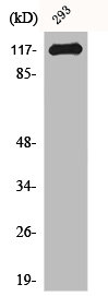
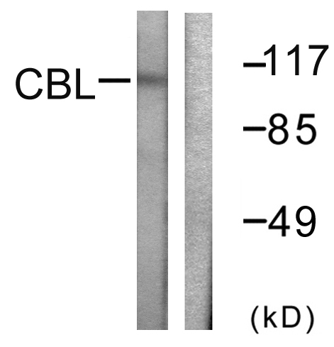
CBL Polyclonal Antibody
RD80126A
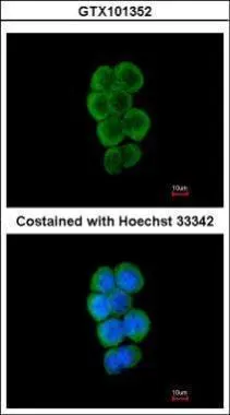
ApplicationsImmunoFluorescence, ImmunoHistoChemistry
Product group Antibodies
ReactivityHuman, Mouse, Rat
TargetCBL
Overview
- SupplierReddot Biotech
- Product NameCBL Polyclonal Antibody
- Delivery Days Customer5
- ApplicationsImmunoFluorescence, ImmunoHistoChemistry
- CertificationResearch Use Only
- Concentration1 mg/ml
- ConjugateUnconjugated
- Gene ID867
- Target nameCBL
- Target descriptionCbl proto-oncogene
- Target synonymsC-CBL, CBL2, FRA11B, NSLL, RNF55, E3 ubiquitin-protein ligase CBL, Cas-Br-M (murine) ecotropic retroviral transforming sequence, Cbl proto-oncogene, E3 ubiquitin protein ligase, RING finger protein 55, RING-type E3 ubiquitin transferase CBL, casitas B-lineage lymphoma proto-oncogene, fragile site, folic acid type, rare, fra(11)(q23.3), oncogene CBL2, proto-oncogene c-Cbl, signal transduction protein CBL
- HostRabbit
- IsotypeIgG
- Protein IDP22681
- Protein NameE3 ubiquitin-protein ligase CBL
- Scientific DescriptionThis gene is a proto-oncogene that encodes a RING finger E3 ubiquitin ligase. The encoded protein is one of the enzymes required for targeting substrates for degradation by the proteasome. This protein mediates the transfer of ubiquitin from ubiquitin conjugating enzymes (E2) to specific substrates. This protein also contains an N-terminal phosphotyrosine binding domain that allows it to interact with numerous tyrosine-phosphorylated substrates and target them for proteasome degradation. As such it functions as a negative regulator of many signal transduction pathways. This gene has been found to be mutated or translocated in many cancers including acute myeloid leukaemia, and expansion of CGG repeats in the 5 UTR has been associated with Jacobsen syndrome. Mutations in this gene are also the cause of Noonan syndrome-like disorder. - This is a CBL Polyclonal Antibody from Reddot Biotech. This product is for Research Use Only.
- ReactivityHuman, Mouse, Rat
- Storage Instruction-20°C
- UNSPSC41116161
Related products
Product group Antibodies
CBL AntibodyCSB-PA001340
ApplicationsImmunoFluorescence, Western Blot, ELISA, ImmunoHistoChemistry
ReactivityHuman, Mouse, Rat
TargetCBL
- SizePrice
Product group Antibodies
CBL Polyclonal AntibodyCAC12849
ApplicationsImmunoFluorescence, Western Blot, ELISA, ImmunoHistoChemistry
TargetCBL
- SizePrice

Product group Antibodies
Anti-CBL Antibody Picoband(r)A00152-2-CARRIER-FREE
ApplicationsFlow Cytometry, Western Blot, ELISA, ImmunoHistoChemistry
ReactivityHuman, Mouse, Rat
TargetCBL
- SizePrice
Product group Antibodies
Anti-CBL Antibody144-61644
ApplicationsImmunoFluorescence, Western Blot, ImmunoHistoChemistry
ReactivityHuman, Mouse, Rat
TargetCBL
- SizePrice
Product group Antibodies
Anti-CBL AntibodyA95490
ApplicationsImmunoFluorescence, Western Blot, ELISA, ImmunoHistoChemistry
ReactivityHuman, Mouse, Rat
- SizePrice

Product group Antibodies
Anti-CBL AntibodyHPA027956
ApplicationsWestern Blot, ImmunoCytoChemistry, ImmunoHistoChemistry
ReactivityHuman, Mouse, Rat
TargetCBL
- SizePrice
Product group Antibodies

c-CBL AntibodyLS-C403014
ApplicationsWestern Blot, ELISA
ReactivityHuman, Mouse
TargetCBL
- SizePrice
Product group Antibodies
CBL antibody [C2C3], C-termGTX101352
ApplicationsImmunoFluorescence, Western Blot, ImmunoCytoChemistry
ReactivityHuman
TargetCBL
- SizePrice