


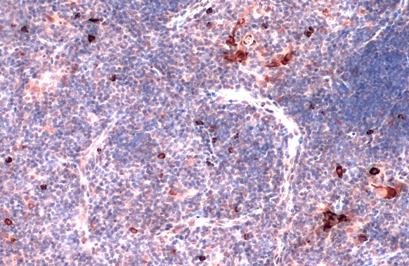
Staining of total mouse peritoneal exudate cells demonstrating labelling of macrophages with Rat anti Mouse CD11b:RPE (GTX76475)
")
Staining of total mouse peritoneal exudate cells demonstrating labelling of macrophages with Rat anti Mouse CD11b:RPE (GTX76475)
CD11b antibody [M1/70.15] (PE)
GTX76470
ApplicationsFlow Cytometry
Product group Antibodies
ReactivityMouse
TargetItgam
Overview
- SupplierGeneTex
- Product NameCD11b antibody [M1/70.15] (PE)
- Delivery Days Customer9
- Application Supplier NoteFor FACS: Use 0.1 ug to label 10^6 cells. Protocols are available upon request. Optimal dilutions/concentrations should be determined by the end user.
- ApplicationsFlow Cytometry
- CertificationResearch Use Only
- ClonalityMonoclonal
- Clone IDM1/70.15
- Concentration0.1 mg/ml
- ConjugateRPE
- Gene ID16409
- Target nameItgam
- Target descriptionintegrin alpha M
- Target synonymsCD11b/CD18, CR3, CR3A, Cd11b, F730045J24Rik, Ly-40, MAC1, Mac-1, Mac-1a, integrin alpha-M, CD11 antigen-like family member B, CD11B (p170), CR-3 alpha chain, Mac-1 alpha, cell surface glycoprotein MAC-1 alpha subunit, cell surface glycoprotein MAC-1 subunit alpha, complement receptor type 3, leukocyte adhesion receptor MO1, macrophage antigen alpha
- HostRat
- IsotypeIgG2b
- Scientific DescriptionRecognises the murine CD11b cell surface antigen (also known as the alpha M integrin chain), a differentiation antigen expressed by granulocytes, monocytes, NK cells and tissue macrophages. The expression of CD11b increases during monocyte maturation and expression levels vary on tissue macrophages. Peritoneal macrophages are reported to express higher levels of CD11b than splenic macrophages. Clone M1/70.15 has been reported to block iC3b binding to its receptor (3).
- ReactivityMouse
- Storage Instruction2°C to 8°C
- UNSPSC41116161
Datasheet
Related products

Product group Antibodies
Anti-CD11B/Integrin Alpha M/Itgam Antibody Picoband(r)A00144-2-CARRIER-FREE
ApplicationsWestern Blot, ELISA
ReactivityMouse
TargetItgam
- SizePrice
Product group Antibodies
CD11b antibodyGTX134493
ApplicationsWestern Blot, ImmunoHistoChemistry, ImmunoHistoChemistry Paraffin
ReactivityMouse, Rat
TargetItgam
- SizePrice
Product group Antibodies
CD11b antibodyGTX134542
ApplicationsWestern Blot, ImmunoHistoChemistry, ImmunoHistoChemistry Paraffin
ReactivityMouse
TargetItgam
- SizePrice
Product group Antibodies
CD11b antibody [M1/70]GTX00608
ApplicationsFlow Cytometry, ImmunoFluorescence, ImmunoPrecipitation, ImmunoCytoChemistry, ImmunoHistoChemistry, ImmunoHistoChemistry Frozen, ImmunoHistoChemistry Paraffin, Neutralisation/Blocking, Other Application
ReactivityHuman, Monkey, Mouse, Primate, Rabbit
TargetItgam
- SizePrice
Product group Antibodies
ApplicationsFlow Cytometry
ReactivityMouse
TargetItgam
- SizePrice
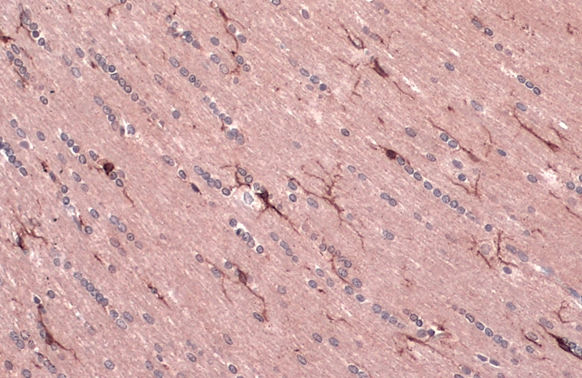
![IHC-Fr analysis of mouse lymph node tissue using GTX76473 CD11b antibody [M1/70.15].](https://www.genetex.com/upload/website/prouct_img/normal/GTX76473/GTX76473_338_IHC-Fr_w_23061322_163.webp)
Product group Antibodies
CD11b antibody [M1/70.15]GTX76473
ApplicationsFlow Cytometry, ImmunoFluorescence, ImmunoPrecipitation, ImmunoCytoChemistry, ImmunoHistoChemistry, ImmunoHistoChemistry Frozen, ImmunoHistoChemistry Paraffin
ReactivityHuman, Mouse, Rabbit
TargetItgam
- SizePrice
Product group Antibodies
CD11b antibody [M1/70.15] (PE)GTX76475
ApplicationsFlow Cytometry
ReactivityHuman, Mouse, Rabbit
TargetItgam
- SizePrice
Product group Antibodies
ApplicationsFunctional Assay, Flow Cytometry, ImmunoPrecipitation, ImmunoHistoChemistry, ImmunoHistoChemistry Frozen
ReactivityHuman, Mouse, Primate, Rabbit
TargetItgam
- SizePrice
![FACS analysis of C57Bl/6 bone marrow cells using GTX80051 CD11b antibody [M1/70] (FITC). Solid line : Primary antibody Dashed line : FITC rat IgG2b isotype control Antibody amount : 0.5 μg](https://www.genetex.com/upload/website/prouct_img/normal/GTX80051/GTX80051_20201020_FACS_27_w_23061322_647.webp)
Product group Antibodies
CD11b antibody [M1/70] (FITC)GTX80051
ApplicationsFlow Cytometry
ReactivityHuman, Monkey, Mouse, Primate
TargetItgam
- SizePrice
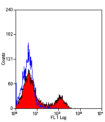
![FACS analysis of C57Bl/6 bone marrow cells using GTX80052 CD11b antibody [M1/70] (PE). Solid line : Primary antibody Dashed line : PE rat IgG2b isotype control Antibody amount : 0.125 μg](https://www.genetex.com/upload/website/prouct_img/normal/GTX80052/GTX80052_20201020_FACS_51_w_23061322_353.webp)
Product group Antibodies
CD11b antibody [M1/70] (PE)GTX80052
ApplicationsFlow Cytometry
ReactivityHuman, Monkey, Mouse, Primate
TargetItgam
- SizePrice