

CD1b(RIV12), CF405S conjugate, 0.1mg/mL [26628-22-8]
BNC040317
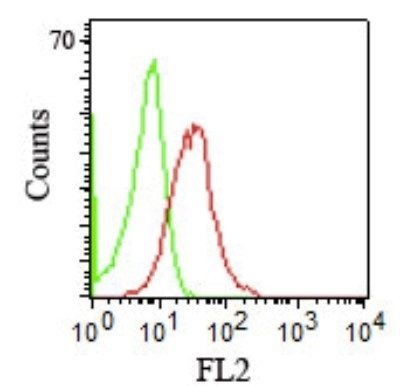
ApplicationsFlow Cytometry
Product group Antibodies
TargetCD1B
Overview
- SupplierBiotium
- Product NameCD1b(RIV12), CF405S conjugate, 0.1mg/mL [26628-22-8]
- Delivery Days Customer9
- ApplicationsFlow Cytometry
- CertificationResearch Use Only
- ClonalityMonoclonal
- Clone IDRIV12
- Concentration0.1 mg/ml
- ConjugateOther Conjugate
- Gene ID910
- Target nameCD1B
- Target descriptionCD1b molecule
- Target synonymsCD1, CD1A, R1, T-cell surface glycoprotein CD1b, CD1B antigen, b polypeptide, cortical thymocyte antigen CD1B, differentiation antigen CD1-alpha-3
- HostMouse
- IsotypeIgG1
- Protein IDP29016
- Protein NameT-cell surface glycoprotein CD1b
- Scientific DescriptionThe mouse monoclonal antibody recognizes CD1b, a 44 kDa type I glycoprotein associated with beta2-microglobulin. It is expressed on dendritic cells, Langerhans cells, thymocytes, and T acute lymphoblastic leukemia cells. The CD1 multigene family encodes five forms of the CD1 T-cell surface glycoprotein in human, designated CD1A, 1B, 1C, 1D and 1E. CD1, a type 1 membrane protein, has structural similarity to the MHC class I antigen and has been shown to present lipid antigens for recognition by T lymphocytes. Constitutive endocytosis of CD1B molecules and the differential sorting of MHC class II from lysosomes separate peptide- and lipid antigen-presenting molecules during dendritic cell maturation. CD1B is also expressed in interdigitating cells.Primary antibodies are available purified, or with a selection of fluorescent CF® Dyes and other labels. CF® Dyes offer exceptional brightness and photostability. Note: Conjugates of blue fluorescent dyes like CF®405S and CF®405M are not recommended for detecting low abundance targets, because blue dyes have lower fluorescence and can give higher non-specific background than other dye colors.
- SourceAnimal
- Storage Instruction2°C to 8°C
- UNSPSC12352203
Related products
Product group Antibodies
CD1b Monoclonal AntibodyBSM-60395M
ApplicationsFlow Cytometry
TargetCD1B
- SizePrice
Product group Antibodies

CD1B AntibodyLS-C829969
ApplicationsELISA, ImmunoHistoChemistry
TargetCD1B
- SizePrice

Product group Antibodies
Anti-CD1B AntibodyHPA021824
ApplicationsWestern Blot, ImmunoHistoChemistry
ReactivityHuman
TargetCD1B
- SizePrice
Product group Antibodies
CD1B AntibodyCSB-PA004890ESR2HU
ApplicationsELISA, ImmunoHistoChemistry
ReactivityHuman
TargetCD1B
- SizePrice

Product group Antibodies
Anti-CD1b Antibody Picoband(r)A02158-1-CARRIER-FREE
ApplicationsWestern Blot, ImmunoHistoChemistry
TargetCD1B
- SizePrice