

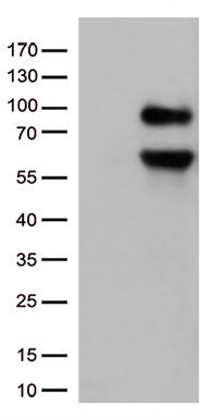
HEK293T cells were transfected with the pCMV6-ENTRY control (Left lane) or pCMV6-ENTRY CD36 (RC203254, Right lane) cDNA for 48 hrs and lysed. Equivalent amounts of cell lysates (5 ug per lane) were separated by SDS-PAGE and immunoblotted with anti-CD36 (1:500).
HEK293T cells were transfected with the pCMV6-ENTRY control (Left lane) or pCMV6-ENTRY CD36 (RC203254, Right lane) cDNA for 48 hrs and lysed. Equivalent amounts of cell lysates (5 ug per lane) were separated by SDS-PAGE and immunoblotted with anti-CD36 (1:500).
CD36 Mouse Monoclonal Antibody [Clone ID: OTI4H7]
CF500935
ApplicationsFlow Cytometry, ImmunoFluorescence, Western Blot, ImmunoHistoChemistry
Product group Antibodies
ReactivityHuman, Rat
TargetCD36
Overview
- SupplierOriGene
- Product NameCD36 Mouse Monoclonal Antibody [Clone ID: OTI4H7]
- Delivery Days Customer14
- ApplicationsFlow Cytometry, ImmunoFluorescence, Western Blot, ImmunoHistoChemistry
- CertificationResearch Use Only
- ClonalityMonoclonal
- Clone IDOTI4H7
- Gene ID948
- Target nameCD36
- Target descriptionCD36 molecule (CD36 blood group)
- Target synonymsBDPLT10, CHDS7, FAT, GP3B, GP4, GPIV, PASIV, SCARB3, platelet glycoprotein 4, CD36 antigen (collagen type I receptor, thrombospondin receptor), CD36 molecule (CD36 blood group) transcript, CD36 molecule (thrombospondin receptor), GPIIIB, PAS IV, PAS-4 protein, cluster determinant 36, fatty acid translocase, glycoprotein IIIb, leukocyte differentiation antigen CD36, platelet glycoprotein IV, scavenger receptor class B, member 3
- HostMouse
- IsotypeIgG1
- Protein IDP16671
- Protein NamePlatelet glycoprotein 4
- Scientific DescriptionCarrier-free (BSA/glycerol-free) CD36 mouse monoclonal antibody, clone OTI4H7 (formerly 4H7)
- ReactivityHuman, Rat
- Storage Instruction-20°C
- UNSPSC12352203
MSDS
Related products
Product group Antibodies
CD36 AntibodyCSB-PA006183
ApplicationsWestern Blot, ELISA
ReactivityHuman, Mouse, Rat
TargetCD36
- SizePrice

Product group Antibodies
References
Anti-CD36 Antibody Picoband(r)A01189-1-CARRIER-FREE
ApplicationsFlow Cytometry, Western Blot
ReactivityHuman, Mouse, Rat
TargetCD36
- SizePrice
Product group Antibodies

Anti-CD36 [185.IG2], Human IgG1, kappaAB04254-10.0
ApplicationsImmunoFluorescence, ImmunoPrecipitation, ImmunoHistoChemistry, Neutralisation/Blocking
ReactivityHuman, Mouse
TargetCD36
- SizePrice
Product group Antibodies
Anti-CD36 AntibodyA98246
ApplicationsWestern Blot, ELISA
ReactivityHuman, Mouse, Rat
- SizePrice
Product group Antibodies

CD36 Antibody (clone 5-271, FITC)LS-C811575
ApplicationsFlow Cytometry
ReactivityHuman
TargetCD36
- SizePrice

Product group Antibodies
Anti-CD36 AntibodyHPA002018
ApplicationsImmunoCytoChemistry, ImmunoHistoChemistry
ReactivityHuman
TargetCD36
- SizePrice
Product group Antibodies
Cd36 Polyclonal AntibodyCAC09109
ApplicationsImmunoFluorescence, Western Blot, ELISA, ImmunoHistoChemistry
ReactivityMouse
TargetCD36
- SizePrice
![Untreated (–) and treated (+) THP-1 whole cell extracts (30 μg) were separated by 7.5% SDS-PAGE, and the membrane was blotted with CD36 antibody [C1C3] (GTX100642) diluted at 1:1000. The HRP-conjugated anti-rabbit IgG antibody (GTX213110-01) was used to detect the primary antibody, and the signal was developed with Trident ECL plus-Enhanced.](https://www.genetex.com/upload/website/prouct_img/normal/GTX100642/GTX100642_42298_20220923_WB_treatment_PMA_22092622_830.webp)
Product group Antibodies
CD36 antibody [C1C3]GTX100642
ApplicationsImmunoFluorescence, Western Blot, ImmunoCytoChemistry, ImmunoHistoChemistry, ImmunoHistoChemistry Paraffin
ReactivityHuman, Mouse
TargetCD36
- SizePrice