

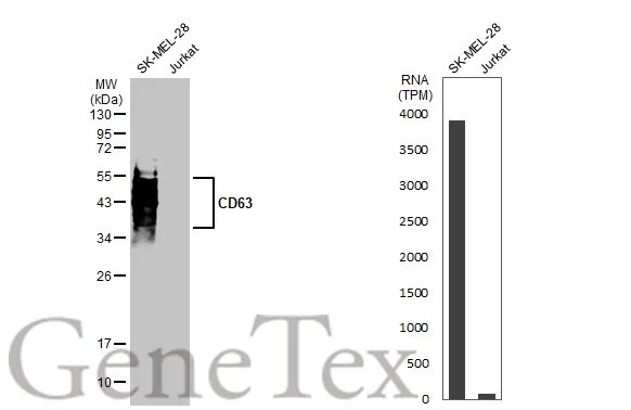
Various whole cell extracts (30 μg) were separated by 12% SDS-PAGE, and the membrane was blotted with CD63 antibody (GTX135220) diluted at 1:1000. The HRP-conjated anti-rabbit IgG antibody (GTX213110-01) was used to detect the primary antibody. Corresponding RNA expression data for the same cell lines are based on Human Protein Atlas program.
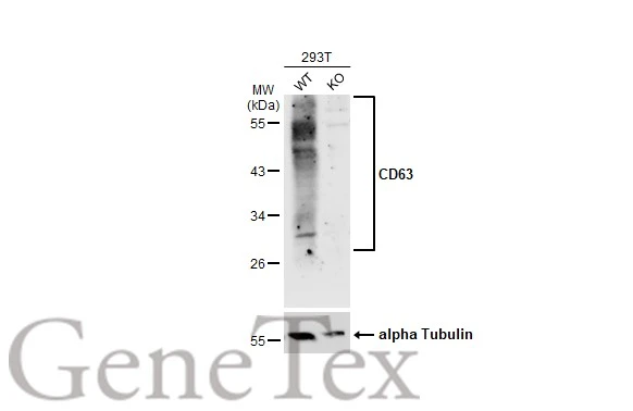
 were separated by 12% SDS-PAGE, and the membrane was blotted with CD63 antibody (GTX135220) diluted at 1:1000. The HRP-conjugated anti-rabbit IgG antibody (GTX213110-01) was used to detect the primary antibody.")
![CD63 antibody detects CD63 protein at cell membrane by immunofluorescent analysis. Sample: HeLa cells were fixed in ice-cold MeOH for 5 min. Green: CD63 stained by CD63 antibody (GTX135220) diluted at 1:1000. Red: alpha Tubulin, a cytoskeleton marker, stained by alpha Tubulin antibody [GT114] (GTX628802) diluted at 1:1000. Blue: Fluoroshield with DAPI (GTX30920).](https://www.genetex.com/upload/website/prouct_img/normal/GTX135220/GTX135220_43957_20211126_ICC_IF_w_23060620_284.webp "CD63 antibody detects CD63 protein at cell membrane by immunofluorescent analysis. Sample: HeLa cells were fixed in ice-cold MeOH for 5 min. Green: CD63 stained by CD63 antibody (GTX135220) diluted at 1:1000. Red: alpha Tubulin, a cytoskeleton marker, stained by alpha Tubulin antibody [GT114] (GTX628802) diluted at 1:1000. Blue: Fluoroshield with DAPI (GTX30920).")
Various whole cell extracts (30 μg) were separated by 12% SDS-PAGE, and the membrane was blotted with CD63 antibody (GTX135220) diluted at 1:1000. The HRP-conjated anti-rabbit IgG antibody (GTX213110-01) was used to detect the primary antibody. Corresponding RNA expression data for the same cell lines are based on Human Protein Atlas program.
CD63 antibody
GTX135220
ApplicationsImmunoFluorescence, Western Blot, ImmunoCytoChemistry
Product group Antibodies
ReactivityHuman
TargetCD63
Overview
- SupplierGeneTex
- Product NameCD63 antibody
- Delivery Days Customer9
- Application Supplier NoteWB: 1:1000. *Optimal dilutions/concentrations should be determined by the researcher.Not tested in other applications.
- ApplicationsImmunoFluorescence, Western Blot, ImmunoCytoChemistry
- CertificationResearch Use Only
- ClonalityPolyclonal
- Concentration1.89 mg/ml
- ConjugateUnconjugated
- Gene ID967
- Target nameCD63
- Target descriptionCD63 molecule
- Target synonymsAD1, HOP-26, ME491, MLA1, OMA81H, Pltgp40, TSPAN30, CD63 antigen, AD1 antigen, CD63 antigen (melanoma 1 antigen), granulophysin, limp1, melanoma-associated antigen ME491, melanoma-associated antigen MLA1, ocular melanoma-associated antigen, tetraspanin-30, tspan-30
- HostRabbit
- IsotypeIgG
- Protein IDP08962
- Protein NameCD63 antigen
- Scientific DescriptionThe protein encoded by this gene is a member of the transmembrane 4 superfamily, also known as the tetraspanin family. Most of these members are cell-surface proteins that are characterized by the presence of four hydrophobic domains. The proteins mediate signal transduction events that play a role in the regulation of cell development, activation, growth and motility. The encoded protein is a cell surface glycoprotein that is known to complex with integrins. It may function as a blood platelet activation marker. Deficiency of this protein is associated with Hermansky-Pudlak syndrome. Also this gene has been associated with tumor progression. Alternative splicing results in multiple transcript variants encoding different protein isoforms. [provided by RefSeq, Apr 2012]
- ReactivityHuman
- Storage Instruction-20°C or -80°C,2°C to 8°C
- UNSPSC41116161
Datasheet
Related products
Product group Antibodies

Anti-CD63 [MOF11]Ab00388-1.1
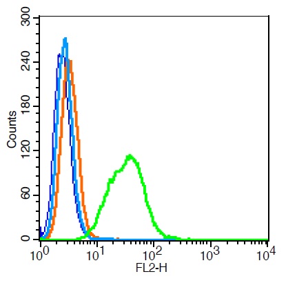
ApplicationsFlow Cytometry
ReactivityHuman
TargetCD63
- SizePrice

Product group Antibodies
Anti-CD63 AntibodyA101276
ApplicationsELISA, ImmunoHistoChemistry
ReactivityHuman
- SizePrice
Product group Antibodies
References
CD63 Polyclonal AntibodyBS-1523R
ApplicationsFlow Cytometry, ImmunoFluorescence, Western Blot, ELISA, ImmunoCytoChemistry, ImmunoHistoChemistry, ImmunoHistoChemistry Frozen, ImmunoHistoChemistry Paraffin
ReactivityHuman
TargetCD63
- SizePrice
Product group Antibodies
CD63 AntibodyCSB-PA006039
ApplicationsWestern Blot, ELISA
ReactivityHuman
TargetCD63
- SizePrice
Product group Antibodies
ApplicationsFlow Cytometry
TargetCD63
- SizePrice
Product group Antibodies

ApplicationsImmunoFluorescence, ELISA, ImmunoHistoChemistry, ImmunoHistoChemistry Frozen, ImmunoHistoChemistry Paraffin
ReactivityHuman
TargetCD63
- SizePrice
Product group Antibodies
CD63 antibodyGTX132953
ApplicationsWestern Blot, ImmunoHistoChemistry, ImmunoHistoChemistry Paraffin
ReactivityHuman
TargetCD63
- SizePrice
Product group Antibodies
References
CD63 antibodyGTX17441
ApplicationsWestern Blot, ImmunoHistoChemistry, ImmunoHistoChemistry Paraffin
ReactivityHuman, Mouse, Rat
TargetCD63
- SizePrice