

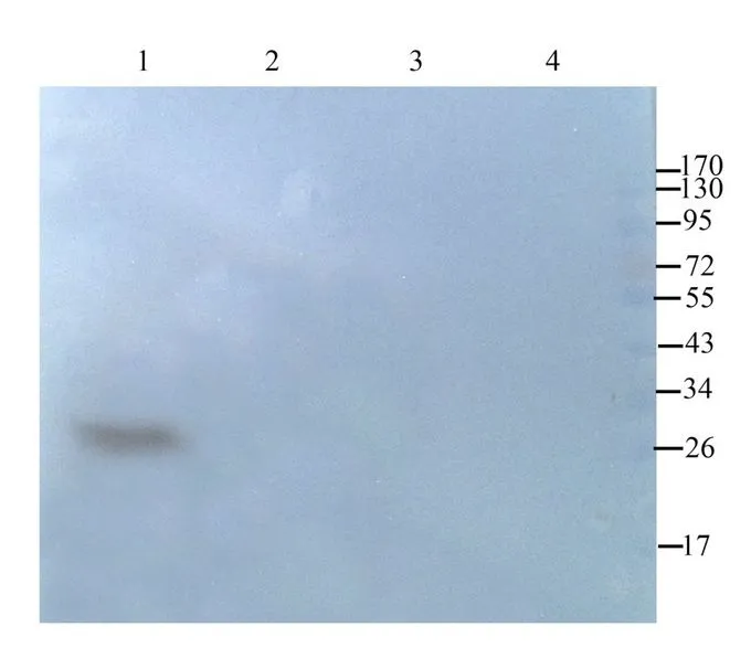
WB analysis of mouse heart (Lane 1), rat kidney (Lane 2), mouse spleen (Lane 3), rat thyroid (Lane 4) lysates using GTX37555 CD63 antibody. Dilution : 1μg/ml


WB analysis of mouse heart (Lane 1), rat kidney (Lane 2), mouse spleen (Lane 3), rat thyroid (Lane 4) lysates using GTX37555 CD63 antibody. Dilution : 1μg/ml
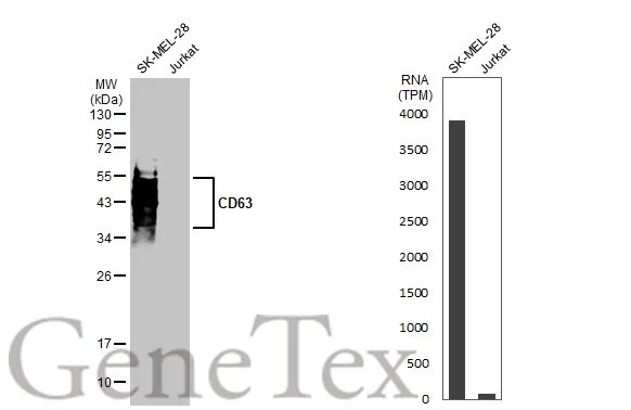
CD63 antibody
GTX37555
ApplicationsWestern Blot, ImmunoHistoChemistry, ImmunoHistoChemistry Paraffin
Product group Antibodies
ReactivityHuman, Mouse, Rat
TargetCD63
Overview
- SupplierGeneTex
- Product NameCD63 antibody
- Delivery Days Customer9
- Application Supplier NoteWB: 1:100-1000. IHC-P: 1:100-500 (based on 0.5 mg/ml). *Optimal dilutions/concentrations should be determined by the researcher.Not tested in other applications.
- ApplicationsWestern Blot, ImmunoHistoChemistry, ImmunoHistoChemistry Paraffin
- CertificationResearch Use Only
- ClonalityPolyclonal
- Concentration0.5 mg/ml
- ConjugateUnconjugated
- Gene ID967
- Target nameCD63
- Target descriptionCD63 molecule
- Target synonymsAD1, HOP-26, ME491, MLA1, OMA81H, Pltgp40, TSPAN30, CD63 antigen, AD1 antigen, CD63 antigen (melanoma 1 antigen), granulophysin, limp1, melanoma-associated antigen ME491, melanoma-associated antigen MLA1, ocular melanoma-associated antigen, tetraspanin-30, tspan-30
- HostRabbit
- IsotypeIgG
- Protein IDP08962
- Protein NameCD63 antigen
- Scientific DescriptionThe protein encoded by this gene is a member of the transmembrane 4 superfamily, also known as the tetraspanin family. Most of these members are cell-surface proteins that are characterized by the presence of four hydrophobic domains. The proteins mediate signal transduction events that play a role in the regulation of cell development, activation, growth and motility. The encoded protein is a cell surface glycoprotein that is known to complex with integrins. It may function as a blood platelet activation marker. Deficiency of this protein is associated with Hermansky-Pudlak syndrome. Also this gene has been associated with tumor progression. Alternative splicing results in multiple transcript variants encoding different protein isoforms. [provided by RefSeq, Apr 2012]
- ReactivityHuman, Mouse, Rat
- Storage Instruction-20°C or -80°C,2°C to 8°C
- UNSPSC41116161
References
- Exosomal miR-27b-3p Derived from Hypoxic Cardiac Microvascular Endothelial Cells Alleviates Rat Myocardial Ischemia/Reperfusion Injury through Inhibiting Oxidative Stress-Induced Pyroptosis via Foxo1/GSDMD Signaling.Read this paper
- Human induced pluripotent stem cell-derived extracellular vesicles reduce hepatic stellate cell activation and liver fibrosis. Povero D et al., 2019 Jun 11, JCI InsightRead this paper
- MiR-21 in extracellular vesicles contributes to the growth of fertilized eggs and embryo development in mice. Lv C et al., 2018 Aug 31, Biosci RepRead this paper
- Large-scale isolation and cytotoxicity of extracellular vesicles derived from activated human natural killer cells. Jong AY et al., 2017, J Extracell VesiclesRead this paper
- Circulating microRNAs, miR-939, miR-595, miR-519d and miR-494, Identify Cirrhotic Patients with HCC. Fornari F et al., 2015, PLoS OneRead this paper
Datasheet
Related products
Product group Antibodies

Anti-CD63 [MOF11]Ab00388-1.1
ApplicationsFlow Cytometry
ReactivityHuman
TargetCD63
- SizePrice

Product group Antibodies
Anti-CD63 AntibodyA101276
ApplicationsELISA, ImmunoHistoChemistry
ReactivityHuman
- SizePrice
Product group Antibodies
References
CD63 Polyclonal AntibodyBS-1523R
ApplicationsFlow Cytometry, ImmunoFluorescence, Western Blot, ELISA, ImmunoCytoChemistry, ImmunoHistoChemistry, ImmunoHistoChemistry Frozen, ImmunoHistoChemistry Paraffin
ReactivityHuman
TargetCD63
- SizePrice
Product group Antibodies
CD63 AntibodyCSB-PA006039
ApplicationsWestern Blot, ELISA
ReactivityHuman
TargetCD63
- SizePrice
Product group Antibodies
ApplicationsFlow Cytometry
TargetCD63
- SizePrice
Product group Antibodies

ApplicationsImmunoFluorescence, ELISA, ImmunoHistoChemistry, ImmunoHistoChemistry Frozen, ImmunoHistoChemistry Paraffin
ReactivityHuman
TargetCD63
- SizePrice
Product group Antibodies
CD63 antibodyGTX132953
ApplicationsWestern Blot, ImmunoHistoChemistry, ImmunoHistoChemistry Paraffin
ReactivityHuman
TargetCD63
- SizePrice
Product group Antibodies
CD63 antibodyGTX135220
ApplicationsImmunoFluorescence, Western Blot, ImmunoCytoChemistry
ReactivityHuman
TargetCD63
- SizePrice