

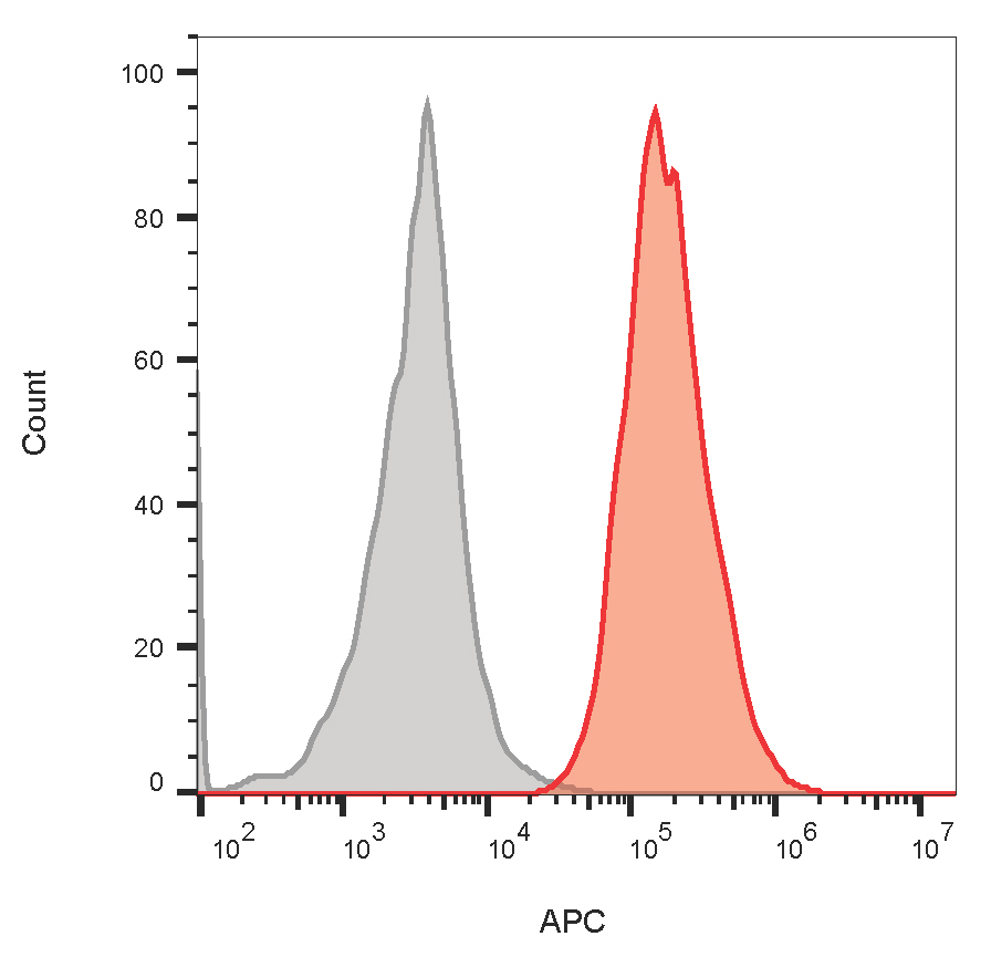
![CD64(10.1), CF647 conjugate, 0.1mg/mL [26628-22-8]](https://biotium.com/wp-content/uploads/2020/10/CD64_101-GAMCF640R-PBMCmono.jpg "CD64(10.1), CF647 conjugate, 0.1mg/mL [26628-22-8]")
CD64(10.1), CF647 conjugate, 0.1mg/mL [26628-22-8]
BNC472846
ApplicationsFunctional Assay, Flow Cytometry, ImmunoFluorescence
Product group Antibodies
TargetFCGR1A
Overview
- SupplierBiotium
- Product NameCD64(10.1), CF647 conjugate, 0.1mg/mL
- Delivery Days Customer9
- ApplicationsFunctional Assay, Flow Cytometry, ImmunoFluorescence
- CertificationResearch Use Only
- ClonalityMonoclonal
- Clone ID10.1
- Concentration0.1 mg/ml
- ConjugateOther Conjugate
- Gene ID2209
- Target nameFCGR1A
- Target descriptionFc fragment of IgG receptor Ia
- Target synonymsCD64; CD64A; Fc fragment of IgG, high affinity Ia, receptor (CD64); Fc fragment of IgG, high affinity Ia, receptor for (CD64); Fc gamma receptor Ia; Fc-gamma receptor I A1; Fc-gamma RI; fc-gamma RIA; FcgammaRI; fcgammaRIa; FCRI; high affinity immunoglobulin gamma Fc receptor I; IGFR1; IgG Fc receptor I
- HostMouse
- IsotypeIgG1
- Protein IDP12314
- Protein NameHigh affinity immunoglobulin gamma Fc receptor I
- Scientific DescriptionCD64 is an Fc receptor that plays a putative role in the initiation of cell-mediated cytotoxicity. Thus far, three genes encoding four distinct CD64 transcripts have been described. CD64 has been shown to associate with signal transducing subunit of the high affinity IgE receptor. Src family kinases Hck and Lyn show increased kinase activity and will co-immunoprecipitate with CD64 subsequent to receptor cross linking. CD64 is constitutively expressed on monocytes and macrophages; exposure of granulocytes to cytokines such as IFN-gamma and G-CSF can induce CD64 expression. Primary antibodies are available purified, or with a selection of fluorescent CF® Dyes and other labels. CF® Dyes offer exceptional brightness and photostability. Note: Conjugates of blue fluorescent dyes like CF®405S and CF®405M are not recommended for detecting low abundance targets, because blue dyes have lower fluorescence and can give higher non-specific background than other dye colors.
- SourceAnimal
- Storage Instruction2°C to 8°C
- UNSPSC12352203
Related products
Product group Antibodies
ApplicationsWestern Blot, ELISA, ImmunoCytoChemistry, ImmunoHistoChemistry, ImmunoHistoChemistry Frozen, ImmunoHistoChemistry Paraffin
TargetFCGR1A
- SizePrice
Product group Antibodies
Anti-FCGR1A AntibodyA29487
ApplicationsImmunoFluorescence, Western Blot, ImmunoHistoChemistry
- SizePrice
Product group Antibodies
ApplicationsFlow Cytometry, ImmunoHistoChemistry
TargetFCGR1A
- SizePrice
Product group Antibodies
Anti-FCGR1A Antibody144-60077
ApplicationsWestern Blot, ImmunoHistoChemistry
TargetFCGR1A
- SizePrice
Product group Antibodies
FCGR1A AntibodyCSB-PA936888
ApplicationsWestern Blot, ELISA, ImmunoHistoChemistry
ReactivityHuman
TargetFCGR1A
- SizePrice
Product group Antibodies

Anti-CD64 [H22]Ab00731-1.4
ApplicationsFlow Cytometry, ELISA, Other Application
TargetFCGR1A
- SizePrice
Product group Antibodies

FCGR1A / CD64 Antibody (clone 10.1)LS-C770018
ApplicationsFlow Cytometry, ImmunoHistoChemistry
TargetFCGR1A
- SizePrice

Product group Antibodies
Anti-FCGR1A Antibody Picoband(r)A02428-1-CARRIER-FREE
ApplicationsFlow Cytometry, Western Blot, ELISA
TargetFCGR1A
- SizePrice
Product group Antibodies
anti-CD64 (human), mAb (10.1) (Fab) (Biotin)ANC-216-530
ApplicationsFlow Cytometry, Western Blot, Neutralisation/Blocking
TargetFCGR1A
- SizePrice