

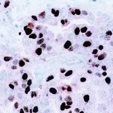
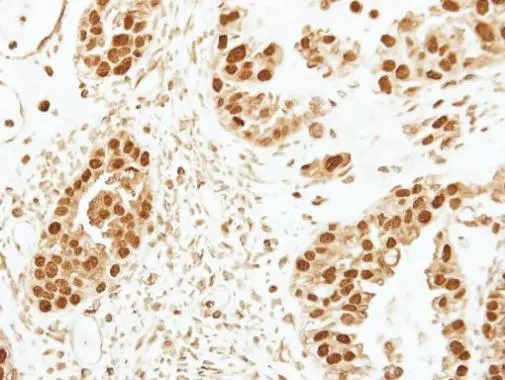
High magnification photomicrograph of an NG3 tumor with a high percentage of mitosin-positive nuclei, indicative of high proliferative activity.
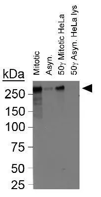
 at 1:500 dilution. Cells were pre-extracted with ice-cold CSK buffer (0.5% Triton X-100, 300 mM Sucrose, 10 mM PIPES, pH8.0) for 4 min, followed by fixation with 4% PFA and regular IFA procedure. Blue channel (DAPI staining) shows chromosomes.")
High magnification photomicrograph of an NG3 tumor with a high percentage of mitosin-positive nuclei, indicative of high proliferative activity.
CENPF antibody [7F11.2]
GTX77645
ApplicationsImmunoFluorescence, ImmunoCytoChemistry, ImmunoHistoChemistry
Product group Antibodies
ReactivityHuman
TargetCENPF
Overview
- SupplierGeneTex
- Product NameCENPF antibody [7F11.2]
- Delivery Days Customer9
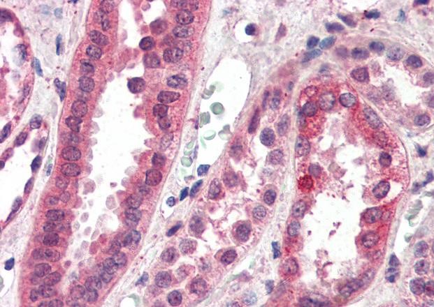
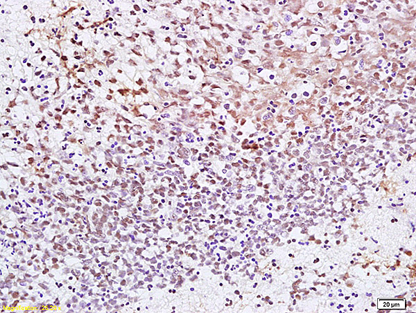
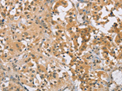
- Application Supplier NoteFor IHC: Use at a concentration of 10 g/mL. Briefly, 4-m tissue sections were cut from the same blocks of hematoxylin and eosin-stained slides, mounted on charged slides, deparaffinized in xylene, and rehydrated in descending grades of ethyl alcohol (from 100% to 70%). The slides used for mitosin staining were then subjected to heat-induced antigen retrieval by immersion in a 0.01 mol/L citrate buffer, pH 6.0; preheated to > 90 C; and heated in an electric vegetable steamer (Black and Decker, Shelton, CT) for 54 minutes. The endogenous peroxidase activity was blocked by a 5-minute treatment with 3% hydrogen peroxide in absolute methanol. Immunohistochemical analysis was then performed using the avidin-biotin technique (LSAB2 peroxidase kit; DAKO, Carpinteria, CA). The antigen-antibody immunoreaction was visualized using 3-3-diaminobenzedine as the chromogen, and the slides were counterstained with Mayer hematoxylin. For ICC/IF: Use at a dilution of 1:500. Optimal dilutions/concentrations should be determined by the researcher.
- ApplicationsImmunoFluorescence, ImmunoCytoChemistry, ImmunoHistoChemistry
- CertificationResearch Use Only
- ClonalityMonoclonal
- Clone ID7F11.2
- Concentration1 mg/ml
- ConjugateUnconjugated
- Gene ID1063
- Target nameCENPF
- Target descriptioncentromere protein F
- Target synonymsCENF, CILD31, PRO1779, STROMS, hcp-1, centromere protein F, AH antigen, CENP-F kinetochore protein, cell-cycle-dependent 350K nuclear protein, centromere protein F, 350/400kDa, kinetochore protein CENPF, mitosin
- HostMouse
- IsotypeIgG2a
- Protein IDP49454
- Protein NameCentromere protein F
- Scientific DescriptionThis gene encodes a protein that associates with the centromere-kinetochore complex. The protein is a component of the nuclear matrix during the G2 phase of interphase. In late G2 the protein associates with the kinetochore and maintains this association through early anaphase. It localizes to the spindle midzone and the intracellular bridge in late anaphase and telophase, respectively, and is thought to be subsequently degraded. The localization of this protein suggests that it may play a role in chromosome segregation during mitotis. It is thought to form either a homodimer or heterodimer. Autoantibodies against this protein have been found in patients with cancer or graft versus host disease. [provided by RefSeq, Jul 2008]
- ReactivityHuman
- Storage Instruction-20°C or -80°C,2°C to 8°C
- UNSPSC41116161
Datasheet
Related products
Product group Antibodies
Anti-CENPF AntibodyA285930
ApplicationsELISA, ImmunoHistoChemistry
ReactivityHuman
- SizePrice

Product group Antibodies
Anti-CENPF Antibody Picoband(r)A03311-1-CARRIER-FREE
ApplicationsFlow Cytometry, ImmunoFluorescence, Western Blot, ELISA, ImmunoCytoChemistry
ReactivityHuman
TargetCENPF
- SizePrice
Product group Antibodies

CENPF / CENP-F AntibodyLS-C832714
ApplicationsELISA, ImmunoHistoChemistry
ReactivityHuman
TargetCENPF
- SizePrice
Product group Antibodies
CENPF Polyclonal AntibodyBS-7839R
ApplicationsImmunoFluorescence, Western Blot, ELISA, ImmunoCytoChemistry, ImmunoHistoChemistry, ImmunoHistoChemistry Frozen, ImmunoHistoChemistry Paraffin
ReactivityHuman
TargetCENPF
- SizePrice
Product group Antibodies

ApplicationsELISA, ImmunoHistoChemistry
ReactivityBovine, Human
TargetCENPF
- SizePrice
Product group Antibodies
ApplicationsWestern Blot, ELISA, ImmunoCytoChemistry, ImmunoHistoChemistry, ImmunoHistoChemistry Frozen, ImmunoHistoChemistry Paraffin
TargetCENPF
- SizePrice
Product group Antibodies
CENPF AntibodyCSB-PA230952
ApplicationsELISA, ImmunoHistoChemistry
ReactivityHuman
TargetCENPF
- SizePrice
Product group Antibodies
References
CENPF antibodyGTX30232
ApplicationsFlow Cytometry, ImmunoFluorescence, ImmunoPrecipitation, Western Blot, ImmunoCytoChemistry
ReactivityBovine, Human
TargetCENPF
- SizePrice
Product group Antibodies
CENPF antibody [C3], C-termGTX100212
ApplicationsImmunoFluorescence, Western Blot, ImmunoCytoChemistry, ImmunoHistoChemistry, ImmunoHistoChemistry Paraffin
ReactivityHuman, Zebra Fish
TargetCENPF
- SizePrice

Product group Antibodies
Anti-CENPF AntibodyHPA052382
ApplicationsImmunoCytoChemistry
ReactivityHuman
TargetCENPF
- SizePrice