

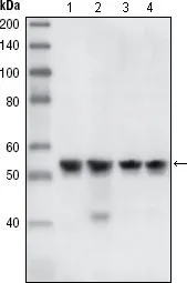
WB analysis of A431 (1), HeLa (2), NIH3T3 (3) and K562 (4) cell lysate using GTX83385 Chk1 antibody [2G1D5].
![WB analysis of HEK293T cells transfected with the pCMV6-ENTRY control (1) and pCMV6-ENTRY CHK1 cDNA (2) using GTX83385 Chk1 antibody [2G1D5].](https://www.genetex.com/upload/website/prouct_img/normal/GTX83385/GTX83385_20170912_WB_1_w_23061322_728.webp "WB analysis of HEK293T cells transfected with the pCMV6-ENTRY control (1) and pCMV6-ENTRY CHK1 cDNA (2) using GTX83385 Chk1 antibody [2G1D5].")
WB analysis of A431 (1), HeLa (2), NIH3T3 (3) and K562 (4) cell lysate using GTX83385 Chk1 antibody [2G1D5].
Chk1 antibody [2G1D5]
GTX83385
ApplicationsImmunoFluorescence, Western Blot, ELISA, ImmunoCytoChemistry
Product group Antibodies
ReactivityHuman, Mouse
TargetCHEK1
Overview
- SupplierGeneTex
- Product NameChk1 antibody [2G1D5]
- Delivery Days Customer7
- Application Supplier NoteWB: 1/500 - 1/2000. ELISA: 1/10000. *Optimal dilutions/concentrations should be determined by the researcher.Not tested in other applications.
- ApplicationsImmunoFluorescence, Western Blot, ELISA, ImmunoCytoChemistry
- CertificationResearch Use Only
- ClonalityMonoclonal
- Clone ID2G1D5
- ConjugateUnconjugated
- Gene ID1111
- Target nameCHEK1
- Target descriptioncheckpoint kinase 1
- Target synonymsCHK1, OZEMA21, serine/threonine-protein kinase Chk1, CHK1 checkpoint homolog, Checkpoint, S. pombe, homolog of, 1, Chk1-S, cell cycle checkpoint kinase
- HostMouse
- IsotypeIgG1
- Protein IDO14757
- Protein NameSerine/threonine-protein kinase Chk1
- Scientific DescriptionThe protein encoded by this gene belongs to the Ser/Thr protein kinase family. It is required for checkpoint mediated cell cycle arrest in response to DNA damage or the presence of unreplicated DNA. This protein acts to integrate signals from ATM and ATR, two cell cycle proteins involved in DNA damage responses, that also associate with chromatin in meiotic prophase I. Phosphorylation of CDC25A protein phosphatase by this protein is required for cells to delay cell cycle progression in response to double-strand DNA breaks. Several alternatively spliced transcript variants have been found for this gene. [provided by RefSeq, Oct 2011]
- ReactivityHuman, Mouse
- Storage Instruction-20°C or -80°C,2°C to 8°C
- UNSPSC12352203
References
- Zhou L, Pei X, Zhang Y, et al. Chk1 Inhibition Potently Blocks STAT3 Tyrosine705 Phosphorylation, DNA-Binding Activity, and Activation of Downstream Targets in Human Multiple Myeloma Cells. Mol Cancer Res. 2022,20(3):456-467. doi: 10.1158/1541-7786.MCR-21-0366Read this paper
Datasheet
Related products
Product group Antibodies
Anti-CHEK1 Antibody144-07653
ApplicationsImmunoFluorescence, Western Blot, ImmunoHistoChemistry
ReactivityHuman, Mouse, Rat
TargetCHEK1
- SizePrice

Product group Antibodies
Anti-Chk1/CHEK1 Antibody Picoband(r)A01060-CARRIER-FREE
ApplicationsFlow Cytometry, Western Blot
ReactivityHuman
TargetCHEK1
- SizePrice
![Chk1 (phospho Ser317) antibody detects Chk1 (phospho Ser317) protein at nucleus by immunofluorescent analysis. Sample: Mock and treated HeLa cells were fixed in 4% paraformaldehyde at RT for 15 min. Green: Chk1 (phospho Ser317) stained by Chk1 (phospho Ser317) antibody (GTX132170) diluted at 1:50. Red: alpha Tubulin, a cytoskeleton marker, stained by alpha Tubulin antibody [GT114] (GTX628802) diluted at 1:1000. Blue: Fluoroshield with DAPI (GTX30920).](https://www.genetex.com/upload/website/prouct_img/normal/GTX132170/GTX132170_44601_20220916_ICC_IF_treatment_UVC_22110201_367.webp)
Product group Antibodies
Chk1 (phospho Ser317) antibodyGTX132170
ApplicationsImmunoFluorescence, Western Blot, ImmunoCytoChemistry
ReactivityHuman
TargetCHEK1
- SizePrice
![Untreated (–) and treated (+) HCT116 whole cell extracts (30 μg) were separated by 10% SDS-PAGE, and the membrane was blotted with Chk1 antibody [ST57-09] (GTX01101) diluted at 1:500. The HRP-conjugated anti-rabbit IgG antibody (GTX213110-01) was used to detect the primary antibody.](https://www.genetex.com/upload/website/prouct_img/normal/GTX01101/GTX01101_HM0401_20220211_WB_treatment_Cisplatin_peptideblocking_w_23053121_692.webp)
Product group Antibodies
Chk1 antibody [ST57-09]GTX01101
ApplicationsWestern Blot
ReactivityHuman
TargetCHEK1
- SizePrice
![Untreated (–) and treated (+) C2C12 whole cell extract (30 μg) were separated by 10% SDS-PAGE, and the membrane was blotted with Chk1 (phospho Ser345) antibody [C1C2], Internal (GTX100065) diluted at 1:1000. The HRP-conjugated anti-rabbit IgG antibody (GTX213110-01) was used to detect the primary antibody, and the signal was developed with Trident ECL plus-Enhanced.](https://www.genetex.com/upload/website/prouct_img/normal/GTX100065/GTX100065_43775_20220701_WB_M_treatment_UVC_22070501_509.webp)
Product group Antibodies
References
ApplicationsImmunoFluorescence, Western Blot, ImmunoCytoChemistry, ImmunoHistoChemistry, ImmunoHistoChemistry Paraffin
ReactivityHuman, Mouse
TargetCHEK1
- SizePrice
Product group Antibodies
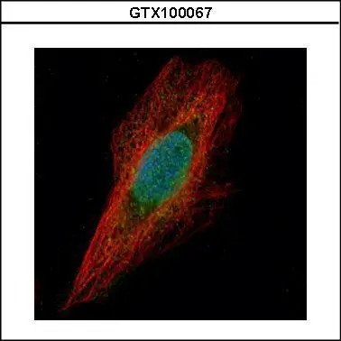
Chk1 antibody [C1C2-3], InternalGTX100067
ApplicationsImmunoFluorescence, ImmunoCytoChemistry
ReactivityHuman
TargetCHEK1
- SizePrice
![CHK1 antibody [C1C2-6], Internal detects Chk1 protein at nucleus by immunofluorescent analysis. Sample: HeLa cells were fixed in 4% paraformaldehyde at RT for 15 min. Green: Chk1 protein stained by CHK1 antibody [C1C2-6], Internal (GTX100070) diluted at 1:500. Red: alpha Tubulin, a cytoskeleton marker, stained by alpha Tubulin antibody [GT114] (GTX628802) diluted at 1:1000. Blue: Hoechst 33342 staining.](https://www.genetex.com/upload/website/prouct_img/normal/GTX100070/GTX100070_39421_20150410_IFA_w_23053123_662.webp)
Product group Antibodies
References
Chk1 antibody [C1C2-6], InternalGTX100070
ApplicationsImmunoFluorescence, Western Blot, ImmunoCytoChemistry, ImmunoHistoChemistry, ImmunoHistoChemistry Paraffin
ReactivityHuman
TargetCHEK1
- SizePrice
![Chk1 (phosphpo Ser345) antibody [HL122] detects Chk1 (phosphpo Ser345) protein at nucleus by immunofluorescent analysis. Sample: Mock and treated HCT116 cells were fixed in 4% paraformaldehyde at RT for 15 min. Green: Chk1 (phosphpo Ser345) stained by Chk1 (phosphpo Ser345) antibody [HL122] (GTX635572) diluted at 1:500. Blue: Fluoroshield with DAPI (GTX30920).](https://www.genetex.com/upload/website/prouct_img/normal/GTX635572/GTX635572_44074_20220325_ICC_IF_treatment_Cisplatin_w_23061202_975.webp)
Product group Antibodies
ApplicationsImmunoFluorescence, Western Blot, ImmunoCytoChemistry
ReactivityHuman
TargetCHEK1
- SizePrice
![Chk1 antibody [6F5] detects Chk1 protein by Western blot analysis. A. 30 μg 293T whole cell lysate/extract B. 30 μg HeLa whole cell lysate/extract C. 30 μg A375 whole cell lysate/extract 10 % SDS-PAGE Chk1 antibody [6F5] (GTX70303) dilution: 1:500](https://www.genetex.com/upload/website/prouct_img/normal/GTX70303/GTX70303_41099_WB_w_23061221_419.webp)
Product group Antibodies
Chk1 antibody [6F5]GTX70303
ApplicationsImmunoFluorescence, Western Blot, ImmunoCytoChemistry
ReactivityHuman
TargetCHEK1
- SizePrice
Product group Antibodies
Chk1 (phospho Ser345) antibodyGTX79111
ApplicationsImmunoHistoChemistry, ImmunoHistoChemistry Paraffin
ReactivityHuman
TargetCHEK1
- SizePrice